File:Mouse blastocyst movie icon.jpg
Mouse_blastocyst_movie_icon.jpg (480 × 480 pixels, file size: 26 KB, MIME type: image/jpeg)
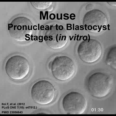
Mouse Blastocyst Development
This shows embryo development observed using time-lapse microscopy from the pronuclear to blastocyst stages without using a specialized incubation chamber.
- Blastocyst Links: MP4 version | Quicktime version | Mouse Development | Blastocyst Development | Movies
Reference
<pubmed>23056643</pubmed>| PLoS ONE
Copyright
© 2012 Itoi et al. This is an open-access article distributed under the terms of the Creative Commons Attribution License, which permits unrestricted use, distribution, and reproduction in any medium, provided the original author and source are credited.
Citation: Itoi F, Tokoro M, Terashita Y, Yamagata K, Fukunaga N, et al. (2012) Offspring from Mouse Embryos Developed Using a Simple Incubator-Free Culture System with a Deoxidizing Agent. PLoS ONE 7(10): e47512. doi:10.1371/journal.pone.0047512
Cite this page: Hill, M.A. (2024, April 19) Embryology Mouse blastocyst movie icon.jpg. Retrieved from https://embryology.med.unsw.edu.au/embryology/index.php/File:Mouse_blastocyst_movie_icon.jpg
- © Dr Mark Hill 2024, UNSW Embryology ISBN: 978 0 7334 2609 4 - UNSW CRICOS Provider Code No. 00098G
File history
Click on a date/time to view the file as it appeared at that time.
| Date/Time | Thumbnail | Dimensions | User | Comment | |
|---|---|---|---|---|---|
| current | 09:57, 14 October 2012 | 480 × 480 (26 KB) | Z8600021 (talk | contribs) | ==Mouse blastocyst development== This shows embryo development observed using time-lapse microscopy from the pronuclear to blastocyst stages without using a specialized incubation chamber. ===Reference=== <pubmed>23056643</pubmed> | [http://www.plosone |
You cannot overwrite this file.
File usage
The following 32 pages use this file:
- Animal Development
- Book - The maturation of the egg of the mouse (1911)
- Lecture - Week 1 and 2 Development
- Models of Human Development
- Morula Development
- Mouse Blastocyst Movie
- Mouse Development
- Mouse E13 microCT Movie
- Mouse Estrous Cycle
- Mouse Heart
- Mouse Stages
- Mouse Timeline
- Mouse Timeline Detailed
- Movies
- Paper - Development of the Mouse Gonads 1
- Paper - Development of the Mouse Gonads 2
- Paper - Development of the Mouse Gonads 3
- Paper - Development of the Mouse Gonads 4
- Paper - Oogenesis in the white mouse (1917)
- Paper - The development of the ear-bones in the mouse
- Paper - The involution of the transitory cortex of the mouse suprarenal (1933)
- Paper - The oestrous cycle in the mouse (1922)
- Paper - The prenatal growth of the mouse
- Quicktime Mouse Blastocyst Movie
- Week 1
- Zygote
- Talk:Quicktime Mouse Blastocyst Movie
- Template:Mouse Blastocyst movie
- Template:Mouse links
- Category:Mouse
- Category:Mouse E12.5
- Category:Mouse E14.5