File:Mouse CT E10.5 head 01.jpg

Original file (1,000 × 636 pixels, file size: 154 KB, MIME type: image/jpeg)
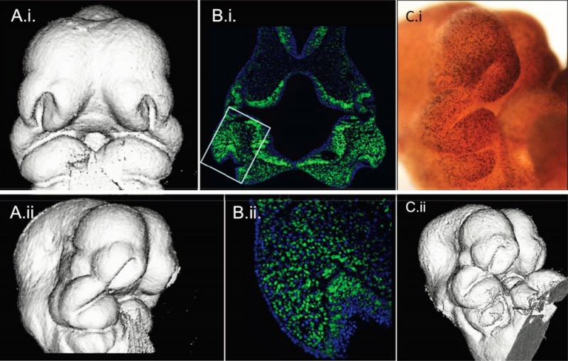
Mouse E10.5 microCT (μCT) renderings and cell proliferation data from the same specimens.
C57BL/J strain
A, 3D reconstruction of μCT taken after processing but before sectioning shown in anterior and right lateral 3/4 views.
B. i, ii Hoescht 33342 staining to visualize cell nuclei (blue) with cells in S-phase visualized using EdU + Alexa Fluor® 488 labeling (green) in frontal sections at the level of the maxillary prominence. B.i at 50× and B.ii and 200×.
Small box in B.i. shows the region magnified below.
C.ii, μCT rendering and same specimen (C.i) processed wholemount for anti-PHH3 primary antibody to identify M-phase cells shown in right lateral 3/4 view.
Figure 9 - http://www.ncbi.nlm.nih.gov/pmc/articles/PMC2836989/figure/F9/
Lab website - http://homepages.ucalgary.ca/%7Emorpho/index.html
Micro-computed tomography-based phenotypic approaches in embryology: procedural artifacts on assessments of embryonic craniofacial growth and development. Schmidt EJ, Parsons TE, Jamniczky HA, Gitelman J, Trpkov C, Boughner JC, Logan CC, Sensen CW, Hallgrímsson B. BMC Dev Biol. 2010 Feb 17;10:18. PMID: 20163731
BMC Dev Biol. 2010; 10: 18.
Published online 2010 February 17. doi: 10.1186/1471-213X-10-18.
PMCID: PMC2836989
Copyright ©2010 Schmidt et al; licensee BioMed Central Ltd.
This is an Open Access article distributed under the terms of the Creative Commons Attribution License (http://creativecommons.org/licenses/by/2.0), which permits unrestricted use, distribution, and reproduction in any medium, provided the original work is properly cited.
File history
Click on a date/time to view the file as it appeared at that time.
| Date/Time | Thumbnail | Dimensions | User | Comment | |
|---|---|---|---|---|---|
| current | 02:05, 16 April 2010 | | 1,000 × 636 (154 KB) | S8600021 (talk | contribs) | Mouse E10.5 microCT (μCT) renderings and cell proliferation data from the same specimens. C57BL/J strain A, 3D reconstruction of μCT taken after processing but before sectioning shown in anterior and right lateral 3/4 views. B. i, ii Hoescht 33342 |
You cannot overwrite this file.
File usage
There are no pages that use this file.