File:Mouse - inner ear cartoon.jpg
Mouse_-_inner_ear_cartoon.jpg (800 × 498 pixels, file size: 77 KB, MIME type: image/jpeg)
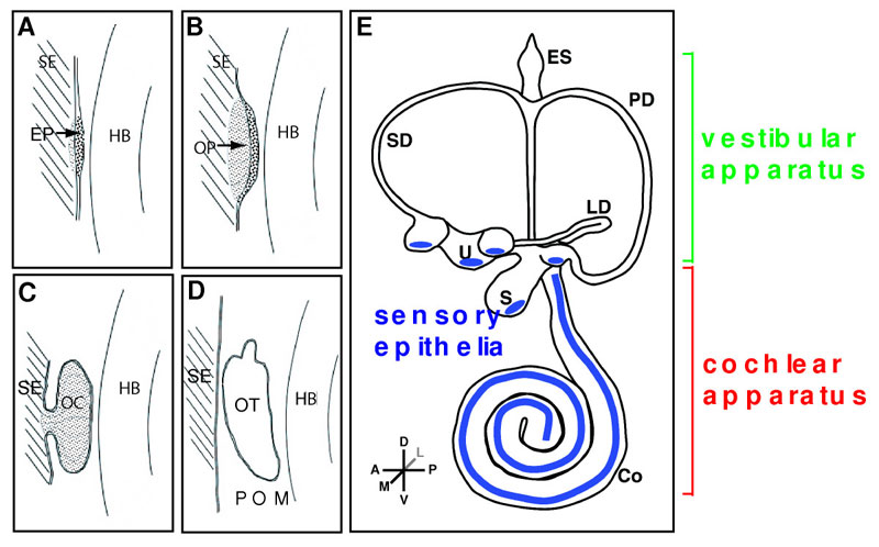
Developmental milestones in mouse inner ear formation
Competence of surface ectoderm lateral to both sides of the hindbrain (HB) precedes any cell morphology changes.
(A) Thickening of surface ectoderm (SE) to form the early placodes (EP) which is primarily driven by Fgf, Wnt and Pax genes.
(B) Invagination of the otic placodes to form the otic pit (OP).
(C) Further development and invagination of the otic pit to form the otic cup (OC) which pinches off from the surface ectoderm.
(D) The separation from the overlying ectoderm gives rise to the otocyst (OT).
(E) Subsequent morphogenesis to finalize the complex 3-dimensional labyrinth which is demarcated into vestibular and cochlear components. Sensory epithelia are shown in blue.
Abbreviations:
- Co - cochlea
- ES - endolymphatic sac
- HB - hindbrain
- LD - lateral semicircular duct
- PD - posterior semicircular duct
- POM - periotic mesenchyme
- S - saccule
- SD - superior semicircular duct
- SE - surface ectoderm
- U - utricle
Reference
<pubmed>20637105</pubmed>| PMC2915946 | BMC Genet.
Chatterjee et al. BMC Genetics 2010 11:68 doi:10.1186/1471-2156-11-68
© 2010 Chatterjee et al; licensee BioMed Central Ltd. This is an Open Access article distributed under the terms of the Creative Commons Attribution License (http://creativecommons.org/licenses/by/2.0), which permits unrestricted use, distribution, and reproduction in any medium, provided the original work is properly cited.
File history
Click on a date/time to view the file as it appeared at that time.
| Date/Time | Thumbnail | Dimensions | User | Comment | |
|---|---|---|---|---|---|
| current | 17:45, 17 November 2010 | | 800 × 498 (77 KB) | S8600021 (talk | contribs) | ==Developmental milestones in mouse inner ear formation== Competence of surface ectoderm lateral to both sides of the hindbrain (HB) precedes any cell morphology changes. (A) Thickening of surface ectoderm (SE) to form the early placodes (EP) which is |
You cannot overwrite this file.
File usage
The following 2 pages use this file: