File:Mouse-morula 01.jpg
Mouse-morula_01.jpg (400 × 398 pixels, file size: 15 KB, MIME type: image/jpeg)
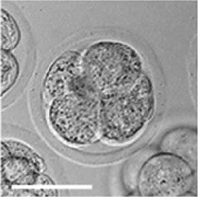
Mouse Morula
Solid ball of cells formed by early mitotic cell division.
Scale bar 50 µm.
- Image Links: zygote | blastomeres | morula | early blastocyst | hatched blastocyst | Mouse Development | Fertilization | Morula | Blastocyst
Reference
<pubmed>20405021</pubmed>| PMC2854157 | PLoS
Editor: Mai Har Sham, The University of Hong Kong, China
Received: December 3, 2009; Accepted: March 23, 2010; Published: April 13, 2010
Copyright: © 2010 Valley et al. This is an open-access article distributed under the terms of the Creative Commons Attribution License, which permits unrestricted use, distribution, and reproduction in any medium, provided the original author and source are credited.
Original file name: Figure 3 Viability of Embryos Post-assay. (image extracted from full figure)
File history
Click on a date/time to view the file as it appeared at that time.
| Date/Time | Thumbnail | Dimensions | User | Comment | |
|---|---|---|---|---|---|
| current | 13:35, 17 April 2010 | | 400 × 398 (15 KB) | S8600021 (talk | contribs) |
You cannot overwrite this file.
File usage
The following 3 pages use this file: