File:Monocyte 01.jpg
From Embryology
No higher resolution available.
Monocyte_01.jpg (480 × 600 pixels, file size: 39 KB, MIME type: image/jpeg)
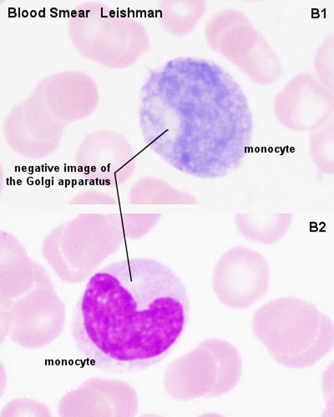
Monocyte
- slightly larger than granulocytes (about 12-18 µm in diameter).
- cytoplasm stains usually stronger than that of granulocytes.
- does not contain any structures which would be visible in the light microscope using most traditional stains (a few very fine bluish gains may be visible in some monocytes).
- has a C-shaped nucleus.
- granules (visible in the EM) correspond to the primary granules of neutrophils (lysosomes).
- Blood Histology: Blood Development | Blood Cell Number Table | Lymphocyte 1 | Lymphocyte 2 | Lymphocyte 3 | Lymphocyte 4 | Monocyte 1 | Monocyte 2 | Monocyte 3 | Monocyte 4 | Neutrophils 1 | Neutrophil 2 | Neutrophil 3 | Neutrophil 4 | Eosinophil 1 | Eosinophil 2 | labeled Neutrophil and Eosinophil | unlabeled - Neutrophil and Eosinophil | Basophil 1 | Basophil 2 | Basophil 3 | Platelet 1 | Platelet 2 | Reticulocyte | Megakaryocyte | Movie | Bone Marrow Histology | Category:Blood
| Blood Cells |
|---|
Adult human blood cell numbers shown in the table below is for reference purposes.
Blood Cell NumbersThe adult ranges of cells / 1 litre (l), total blood volume is about 4.7 to 5 litres. Blood Development | Blood Histology Red Blood Cells
Leukocytes (white blood cells)
Granulocytes
Non-Granulocytes
Lymphocytes
Platelets
|
Links: Histology | Histology Stains | Blue Histology images copyright Lutz Slomianka 1998-2009. The literary and artistic works on the original Blue Histology website may be reproduced, adapted, published and distributed for non-commercial purposes. See also the page Histology Stains.
Cite this page: Hill, M.A. (2024, April 18) Embryology Monocyte 01.jpg. Retrieved from https://embryology.med.unsw.edu.au/embryology/index.php/File:Monocyte_01.jpg
- © Dr Mark Hill 2024, UNSW Embryology ISBN: 978 0 7334 2609 4 - UNSW CRICOS Provider Code No. 00098G
File history
Click on a date/time to view the file as it appeared at that time.
| Date/Time | Thumbnail | Dimensions | User | Comment | |
|---|---|---|---|---|---|
| current | 08:28, 25 February 2012 | | 480 × 600 (39 KB) | Z8600021 (talk | contribs) | |
| 09:32, 22 December 2010 | 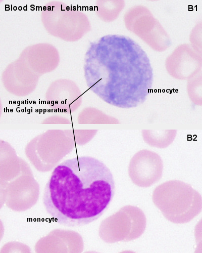 | 400 × 500 (58 KB) | S8600021 (talk | contribs) | Source: Blue Histology - Blood Mono101le.jpg http://www.lab.anhb.uwa.edu.au/mb140/CorePages/Blood/blood.htm |
You cannot overwrite this file.