File:Moffatt1957 plate01.jpg: Difference between revisions
mNo edit summary |
mNo edit summary |
||
| Line 6: | Line 6: | ||
'''Fig. 2''' - Stage {{CS23}} (no. {{CE86}}, 30 mm) shows the further development of the articular disk. In frontal section the upper surface of the developing condyle faces laterally. Lying against this surface is a dark band of cells which gives rise to the articular disk. The external pterygoid muscle joins the medial side of the condyle at its approximate site of attachment in the fully developed joint. The muscle does not end at this point, however. It passes posteriorly along the medial side of the condyle and joins the band of cells forming the articular disk. The cells giving rise to the articular disk and to the external pterygoid tendon continue from this point postero-superiorly to attach to that portion of Meckel’s cartilage which becomes the malleus. This attachment of the mesenchymal layer and the external pterygoid tendon to the malleus is a constant feature, seen in all specimens up to and including a 40mm fetus. In this 30-mm embryo, membrane-bone spicules make their appearance in the zygomatic process and squama of the temporal bone. The articular disk is still separated from the temporal bone by a large area of sparse cells which are giving up their round form to become elongated. The joint capsule is not yet recognizable. | '''Fig. 2''' - Stage {{CS23}} (no. {{CE86}}, 30 mm) shows the further development of the articular disk. In frontal section the upper surface of the developing condyle faces laterally. Lying against this surface is a dark band of cells which gives rise to the articular disk. The external pterygoid muscle joins the medial side of the condyle at its approximate site of attachment in the fully developed joint. The muscle does not end at this point, however. It passes posteriorly along the medial side of the condyle and joins the band of cells forming the articular disk. The cells giving rise to the articular disk and to the external pterygoid tendon continue from this point postero-superiorly to attach to that portion of Meckel’s cartilage which becomes the malleus. This attachment of the mesenchymal layer and the external pterygoid tendon to the malleus is a constant feature, seen in all specimens up to and including a 40mm fetus. In this 30-mm embryo, membrane-bone spicules make their appearance in the zygomatic process and squama of the temporal bone. The articular disk is still separated from the temporal bone by a large area of sparse cells which are giving up their round form to become elongated. The joint capsule is not yet recognizable. | ||
'''Fig. 3''' - [[ | '''Fig. 3''' - [[Fetus]] (no. {{CE1318}}, 37 mm) Relations to adjacent structures. In a frontal section the condyle is still merely a condensation of mesenchyme around the upper end of the osseous lamella forming the mandible. The articular disk and external pterygoid tendon are shown continuing posteriorly to their attachment on the malleus. The structures in immediate relation to the medial side of the temporomandibular joint are demonstrated in this section. Directly inferior to the external pterygoid muscle is the internal maxillary artery cut in cross section. The middle meningeal artery is seen in longitudinal section running superiorly between the condyle and the otic ganglion. The two portions of the auriculo-temporal nerve which surround the middle meningeal artery are visible, as are also the inferior alveolar and lingual nerves. | ||
'''Fig. 4''' - [[Fetal]] (no. {{CE95}}, 46 mm) First appearance of the condylar cartilage. No further development of the joint components was noticed until the condylar cartilage appeared. The cartilage is located at the upper end of the osseous lamella, which forms the posterior border of the mandible. In sagittal section (fig. 4, pl. 1), the growth of this cartilage appears to be directed superiorly and somewhat anteriorly. The condylar cartilage has not grown sufficiently to force the disk into contact with the superior articular element. Immediately above the condyle, between it and the disk, is the external pterygoid tendon cut almost in cross section as it curves laterally to attach to the malleus. The auriculotemporal nerve has been cut obliquely, passing between Mcckel's cartilage and the posterior surface of the condyle. | '''Fig. 4''' - [[Fetal]] (no. {{CE95}}, 46 mm) First appearance of the condylar cartilage. No further development of the joint components was noticed until the condylar cartilage appeared. The cartilage is located at the upper end of the osseous lamella, which forms the posterior border of the mandible. In sagittal section (fig. 4, pl. 1), the growth of this cartilage appears to be directed superiorly and somewhat anteriorly. The condylar cartilage has not grown sufficiently to force the disk into contact with the superior articular element. Immediately above the condyle, between it and the disk, is the external pterygoid tendon cut almost in cross section as it curves laterally to attach to the malleus. The auriculotemporal nerve has been cut obliquely, passing between Mcckel's cartilage and the posterior surface of the condyle. | ||
Revision as of 12:21, 28 October 2018
Plate 1
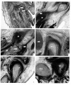
Fig. 1 - Stage 22 embryo (no. 6701, 24 mm) Origin of articular disk. The origin of the articular disk is associated with the appearance of the external pterygoid and masseter muscles, which are recognizable in a frontal section (fig. I, pl. 1) through the region corresponding to the anterior portion of the future condyle, the disk is seen as a vague layer of mesenchyme extending laterally across the superior border of the external pterygoid muscle to the medial side of the masseter. The developing disk is separated from the condensation of mesenchyme which becomes the zygomatic process of the temporal bone by a large area of sparse cells, the precursor of the superior joint cavity. In contrast, the inferior joint cavity is preceded by only a very narrow zone of cells, because the articular disk, even in its early development, rests closely on the mandible. In this specimen an attachment of the external pterygoid muscle to the mandible could not be identified. The muscle, however, could be followed back to the posterior end of Meckel’s cartilage.
Fig. 2 - Stage 23 (no. 86, 30 mm) shows the further development of the articular disk. In frontal section the upper surface of the developing condyle faces laterally. Lying against this surface is a dark band of cells which gives rise to the articular disk. The external pterygoid muscle joins the medial side of the condyle at its approximate site of attachment in the fully developed joint. The muscle does not end at this point, however. It passes posteriorly along the medial side of the condyle and joins the band of cells forming the articular disk. The cells giving rise to the articular disk and to the external pterygoid tendon continue from this point postero-superiorly to attach to that portion of Meckel’s cartilage which becomes the malleus. This attachment of the mesenchymal layer and the external pterygoid tendon to the malleus is a constant feature, seen in all specimens up to and including a 40mm fetus. In this 30-mm embryo, membrane-bone spicules make their appearance in the zygomatic process and squama of the temporal bone. The articular disk is still separated from the temporal bone by a large area of sparse cells which are giving up their round form to become elongated. The joint capsule is not yet recognizable.
Fig. 3 - Fetus (no. 1318, 37 mm) Relations to adjacent structures. In a frontal section the condyle is still merely a condensation of mesenchyme around the upper end of the osseous lamella forming the mandible. The articular disk and external pterygoid tendon are shown continuing posteriorly to their attachment on the malleus. The structures in immediate relation to the medial side of the temporomandibular joint are demonstrated in this section. Directly inferior to the external pterygoid muscle is the internal maxillary artery cut in cross section. The middle meningeal artery is seen in longitudinal section running superiorly between the condyle and the otic ganglion. The two portions of the auriculo-temporal nerve which surround the middle meningeal artery are visible, as are also the inferior alveolar and lingual nerves.
Fig. 4 - Fetal (no. 95, 46 mm) First appearance of the condylar cartilage. No further development of the joint components was noticed until the condylar cartilage appeared. The cartilage is located at the upper end of the osseous lamella, which forms the posterior border of the mandible. In sagittal section (fig. 4, pl. 1), the growth of this cartilage appears to be directed superiorly and somewhat anteriorly. The condylar cartilage has not grown sufficiently to force the disk into contact with the superior articular element. Immediately above the condyle, between it and the disk, is the external pterygoid tendon cut almost in cross section as it curves laterally to attach to the malleus. The auriculotemporal nerve has been cut obliquely, passing between Mcckel's cartilage and the posterior surface of the condyle.
Fig. 5 - Fetal (no. 5652, 49 mm) The articular disk appears to be elevated from the condyle because this section is close to the external pterygoid muscle, which comes between the mesenchymal portion of the disk and the medial part of the condyle. The relationship is better understood by referring to the frontal section. The articular disk lies directly on the condyle except in the area occupied by the external pterygoid tendon. This frontal section illustrates several important points about the articular disk.
Fig. 6 - Fetal (no. 96, 50 mm) The actual passage of the external pterygoid tendon from the condyle to the malleus can be seen in a sagittal section of the temporomandibular joint. The tendon runs obliquely from the upper posterior part of the condyle to the posterior surface of Meckel’s cartilage. In adjacent sections Meckel’s cartilage changes its direction and position slightly so that the pterygoid tendon is actually attached to the lateral side of the malleus. A short segment of the articular disk extends posteriorly above the upper end of the external pterygoid tendon and parallel to the membrane bone that forms the zygomatic process of the temporal bone. This part of the disk when followed posteriorly joins the external pterygoid tendon and travels in common with it to the malleus.
| Historic Disclaimer - information about historic embryology pages |
|---|
| Pages where the terms "Historic" (textbooks, papers, people, recommendations) appear on this site, and sections within pages where this disclaimer appears, indicate that the content and scientific understanding are specific to the time of publication. This means that while some scientific descriptions are still accurate, the terminology and interpretation of the developmental mechanisms reflect the understanding at the time of original publication and those of the preceding periods, these terms, interpretations and recommendations may not reflect our current scientific understanding. (More? Embryology History | Historic Embryology Papers) |
Reference
Moffatt BC. The prenatal development of the human temporomandibular joint. (1957) Carnegie Instn. Wash. Publ. 611, Contrib. Embryol., 36: .
Cite this page: Hill, M.A. (2024, April 19) Embryology Moffatt1957 plate01.jpg. Retrieved from https://embryology.med.unsw.edu.au/embryology/index.php/File:Moffatt1957_plate01.jpg
- © Dr Mark Hill 2024, UNSW Embryology ISBN: 978 0 7334 2609 4 - UNSW CRICOS Provider Code No. 00098G
File history
Click on a date/time to view the file as it appeared at that time.
| Date/Time | Thumbnail | Dimensions | User | Comment | |
|---|---|---|---|---|---|
| current | 18:51, 30 October 2016 |  | 1,500 × 1,803 (878 KB) | Z8600021 (talk | contribs) | {{Historic Disclaimer}} ===Reference=== {{Ref-Moffatt1957}} |
You cannot overwrite this file.
File usage
The following page uses this file:
