File:Mitosis fl.jpg: Difference between revisions
From Embryology
(Rieder's research team uses fluorescent dyes to label the dividing newt lung cells. The scientists use newt lung cells in their studies because these cells are large, easy to see into, and are biochemically similar to human lung cells. Photo: Conly Riede) |
No edit summary |
||
| Line 6: | Line 6: | ||
http://publications.nigms.nih.gov/moleculestomeds/biology.html | http://publications.nigms.nih.gov/moleculestomeds/biology.html | ||
[[Category:Mitosis]] | |||
Revision as of 16:36, 17 November 2009
Rieder's research team uses fluorescent dyes to label the dividing newt lung cells. The scientists use newt lung cells in their studies because these cells are large, easy to see into, and are biochemically similar to human lung cells.
Photo: Conly Rieder
Source: From Molecules to Medicines, National Institute of General Medical Sciences, USA
http://publications.nigms.nih.gov/moleculestomeds/biology.html
File history
Click on a date/time to view the file as it appeared at that time.
| Date/Time | Thumbnail | Dimensions | User | Comment | |
|---|---|---|---|---|---|
| current | 15:25, 27 July 2009 | 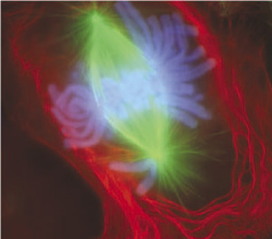 | 300 × 264 (21 KB) | MarkHill (talk | contribs) | Rieder's research team uses fluorescent dyes to label the dividing newt lung cells. The scientists use newt lung cells in their studies because these cells are large, easy to see into, and are biochemically similar to human lung cells. Photo: Conly Riede |
You cannot overwrite this file.
File usage
The following 6 pages use this file: