File:Minot1897 fig006.jpg
From Embryology

No higher resolution available.
Minot1897_fig006.jpg (573 × 503 pixels, file size: 69 KB, MIME type: image/jpeg)
Fig. 6. Ovum of White Mouse. Conjugation of the Pro-nuclei, and Formation of the Segmentation Spindle X 1500 diams. (after Sobotta)
- Chapter II. The Early Development Of Mammals: 1. Human Spermatozoa | 2. Human Ovum | 3. Worm Ovum | 4. Rabbit Ovum 17 Hours | 5. Mouse Ovum early Pro-nuclei | 6. Mouse Ovum Spindle | 7. Mouse Ovum late Pro-nuclei | 8. Rabbit Ovum 24 Hours | 9. Snail Ovum First Cleavage | Chapter 2 Figures | Figures
| Historic Disclaimer - information about historic embryology pages |
|---|
 |
Reference
Minot CS. Human Embryology. (1897) London: The Macmillan Company.
Cite this page: Hill, M.A. (2024, April 16) Embryology Minot1897 fig006.jpg. Retrieved from https://embryology.med.unsw.edu.au/embryology/index.php/File:Minot1897_fig006.jpg
- © Dr Mark Hill 2024, UNSW Embryology ISBN: 978 0 7334 2609 4 - UNSW CRICOS Provider Code No. 00098G
File history
Click on a date/time to view the file as it appeared at that time.
| Date/Time | Thumbnail | Dimensions | User | Comment | |
|---|---|---|---|---|---|
| current | 08:21, 5 April 2014 | | 573 × 503 (69 KB) | Z8600021 (talk | contribs) | |
| 08:20, 5 April 2014 | 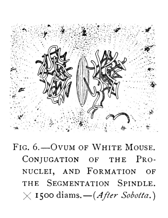 | 590 × 764 (96 KB) | Z8600021 (talk | contribs) | Fig. 6. Ovum of White Mouse. Conjugation of the Pro-nuclei, and Formation of the Segmentation Spindle X 1500 diams. (after Sobotta) {{Minot1897 2 figures}} {{Minot1897 figures}} Category:Mouse |
You cannot overwrite this file.
File usage
The following 2 pages use this file: