File:Microtia.jpg: Difference between revisions
From Embryology
mNo edit summary |
mNo edit summary |
||
| (One intermediate revision by the same user not shown) | |||
| Line 6: | Line 6: | ||
{{Outer ear abnormal links}} | {{Outer ear abnormal links}} | ||
{{Hearing Links}} | |||
===Reference=== | ===Reference=== | ||
NZ National Women's Health [http://www.adhb.govt.nz/newborn/TeachingResources/Dermatology/EarAnomalies.htm Ear Anomalies] | |||
{{Footer}} | |||
[[Category:Senses]] [[Category:Hearing]] [[Category:Abnormal Development]][[Category:Outer Ear]] | [[Category:Senses]] [[Category:Hearing]] [[Category:Abnormal Development]][[Category:Outer Ear]] | ||
Latest revision as of 08:07, 7 April 2016
Newborn Microtia
Microtia refers to an underdeveloped external ear (pinna).
Reference
NZ National Women's Health Ear Anomalies
Cite this page: Hill, M.A. (2024, April 23) Embryology Microtia.jpg. Retrieved from https://embryology.med.unsw.edu.au/embryology/index.php/File:Microtia.jpg
- © Dr Mark Hill 2024, UNSW Embryology ISBN: 978 0 7334 2609 4 - UNSW CRICOS Provider Code No. 00098G
File history
Click on a date/time to view the file as it appeared at that time.
| Date/Time | Thumbnail | Dimensions | User | Comment | |
|---|---|---|---|---|---|
| current | 13:08, 28 September 2009 | 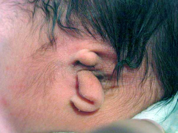 | 600 × 450 (27 KB) | S8600021 (talk | contribs) | Newborn Microtia Image source: NZ National Women's Health [http://www.adhb.govt.nz/newborn/TeachingResources/Dermatology/EarAnomalies.htm Ear Anomalies] |
You cannot overwrite this file.
File usage
The following 12 pages use this file:
- 2009 Lecture 17
- 2010 Lecture 17
- 2011 Lab 10 - Abnormalities
- 2011 Lab 6 - Abnormalities
- ANAT2341 Lab 10 - Abnormalities
- ANAT2341 Lab 6 - Abnormalities
- Abnormal Development - Thalidomide
- BGDB Face and Ear - Abnormalities
- Hearing - Outer Ear Development
- Lecture - Sensory Development
- Sensory - Hearing Abnormalities
- Sensory - Hearing and Balance Development