File:Meyer1920 fig11.jpg: Difference between revisions
mNo edit summary |
|||
| (One intermediate revision by the same user not shown) | |||
| Line 1: | Line 1: | ||
==Fig. | ==Fig. 11.== | ||
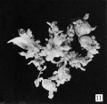
No. 1323 (Dr. J. W. Schlieder) also is a large mass very like the preceding, which completely fills a liter jar. It is accompanied by much clot and composed mainly of a large, thick-walled, hemorrhagic, necrotic mass 80X50X45 mm., containing a large, thin-walled cavity 65X30X25 mm., which is broken at one end. This cavity which is apparently that of the chorionic vesicle, is empty, smooth, and thin-walled, except where it is composed of a characteristic hydatiform mass shown in figure 11. | |||
| Line 5: | Line 7: | ||
{{Meyer1920}} | {{Meyer1920}} | ||
[[Category:Hydatidiform Mole]] | |||
Latest revision as of 23:29, 11 May 2014
Fig. 11.
No. 1323 (Dr. J. W. Schlieder) also is a large mass very like the preceding, which completely fills a liter jar. It is accompanied by much clot and composed mainly of a large, thick-walled, hemorrhagic, necrotic mass 80X50X45 mm., containing a large, thin-walled cavity 65X30X25 mm., which is broken at one end. This cavity which is apparently that of the chorionic vesicle, is empty, smooth, and thin-walled, except where it is composed of a characteristic hydatiform mass shown in figure 11.
Plate 2: Fig. 8 | Fig. 9 | Fig. 10 | Fig. 11 | Fig. 12 | Fig. 13
- Meyer Links: Plate 1 | Plate 2 | Plate 3 | Plate 4 | Plate 5 | Plate 6 | Contribution No.40 | Volume IX | Contributions to Embryology | Hydatidiform Mole | Tubal Pregnancy
| Historic Disclaimer - information about historic embryology pages |
|---|
| Pages where the terms "Historic" (textbooks, papers, people, recommendations) appear on this site, and sections within pages where this disclaimer appears, indicate that the content and scientific understanding are specific to the time of publication. This means that while some scientific descriptions are still accurate, the terminology and interpretation of the developmental mechanisms reflect the understanding at the time of original publication and those of the preceding periods, these terms, interpretations and recommendations may not reflect our current scientific understanding. (More? Embryology History | Historic Embryology Papers) |
Reference
Meyer AW. Hydatiform degeneration in tubal and uterine pregnancy. (1920) Carnegie Instn. Wash. Publ., Contrib. Embryol., 40: 327- 364.
Cite this page: Hill, M.A. (2024, April 18) Embryology Meyer1920 fig11.jpg. Retrieved from https://embryology.med.unsw.edu.au/embryology/index.php/File:Meyer1920_fig11.jpg
- © Dr Mark Hill 2024, UNSW Embryology ISBN: 978 0 7334 2609 4 - UNSW CRICOS Provider Code No. 00098G
File history
Click on a date/time to view the file as it appeared at that time.
| Date/Time | Thumbnail | Dimensions | User | Comment | |
|---|---|---|---|---|---|
| current | 09:58, 8 April 2012 |  | 740 × 719 (39 KB) | Z8600021 (talk | contribs) | ==Fig. 8.== Plate 2: Fig. 8 | Fig. 9 | Fig. 10 | Fig. 11 | Fig. 12 | [[ |
You cannot overwrite this file.
File usage
The following page uses this file:
