File:Mall Meyer1921 plate16.jpg: Difference between revisions
(==Plate 16== Fig. 163. External appearance of intact specimen. No. 1522. X0.75. Fig. 164. External appearance of same specimen, showing where block was removed. X0.75. Fig. 165. Appearance of cross section of pregnant ovary and tube, same specimen. X0.7) |
No edit summary |
||
| Line 26: | Line 26: | ||
Uterine end | Uterine end | ||
Clot containing | Clot containing chiorionic villi Free fimbr. end Site of rupture Posterior view Fig. 161 - | ||
{{Mall Meyer1921Plate16}} | |||
<gallery> | |||
Mall_Meyer1921_fig163.jpg|Fig. 163. External appearance of intact specimen. No. 1522. X0.75. | |||
Mall_Meyer1921_fig164.jpg|Fig. 164. External appearance of same specimen, showing where block was removed. X0.75. | |||
Mall_Meyer1921_fig165.jpg|Fig. 165. Appearance of cross section of pregnant ovary and tube, same specimen. X0.75. | |||
Mall_Meyer1921_fig166.jpg|Fig. 166. Photograph of a section taken from the thick portion of the ovarian stroma near the mesovarium, showing a well-developed Graafian follicle. Same specimen. X2.25. | |||
Mall_Meyer1921_fig167.jpg|Fig. 167. Section of part of the same specimen, showing the clot which contains the empty vesicle largely surrounded by ovarian stroma. XI. 9. | |||
Mall_Meyer1921_fig168.jpg|Fig. 168. Marked clubbing of villi of No. 550. X2.25. | |||
Mall_Meyer1921_fig169.jpg|Fig. 169. Cross section of decidua and conceptus. No. 698. (See Chapter XII.) X4.5. | |||
Mall_Meyer1921_fig170.jpg|Fig. 170. Section of conceptus, decidua, and muscularis. No. 970. (See Chapter XII.) X4.5. | |||
Mall_Meyer1921_fig171.jpg|Fig. 171. Section of decidua and conceptus. No. 962. (See Chapter XII.) X4.5. | |||
Mall_Meyer1921_fig172.jpg|Fig. 172. Section of a part of the conceptus, showing chorionic membrane, cyemic (?) rudiment (x) and yolk-sac. No. 1843. (See Chapter XII.) X51.5. | |||
Mall_Meyer1921_fig173.jpg|Fig. 173. External view of No. 2047, showing the distended amnion. (See Chapter XII.) X2.25. | |||
Mall_Meyer1921_fig174.jpg|Fig. 174. Section of the tube and the conceptus. No. 977. (See Chapter XII.) X3.3. | |||
</gallery> | |||
{{Carnegie56 TOC}} | {{Carnegie56 TOC}} | ||
Latest revision as of 08:38, 13 December 2012
Plate 16
Fig. 163. External appearance of intact specimen. No. 1522. X0.75.
Fig. 164. External appearance of same specimen, showing where block was removed. X0.75.
Fig. 165. Appearance of cross section of pregnant ovary and tube, same specimen. X0.75.
Fig. 166. Photograph of a section taken from the thick portion of the ovarian stroma near the mesovarium, showing a well-developed Graafian follicle. Same specimen. X2.25.
Fig. 167. Section of part of the same specimen, showing the clot which contains the empty vesicle largely surrounded by ovarian stroma. XI. 9.
Fig. 168. Marked clubbing of villi of No. 550. X2.25.
Fig. 169. Cross section of decidua and conceptus. No. 698. (See Chapter XII.) X4.5.
Fig. 170. Section of conceptus, decidua, and muscularis. No. 970. (See Chapter XII.) X4.5.
Fig. 171. Section of decidua and conceptus. No. 962. (See Chapter XII.) X4.5.
Fig. 172. Section of a part of the conceptus, showing chorionic membrane, cyemic (?) rudiment (x) and yolk-sac. No. 1843. (See Chapter XII.) X51.5.
Fig. 173. External view of No. 2047, showing the distended amnion. (See Chapter XII.) X2.25.
Fig. 174. Section of the tube and the conceptus. No. 977. (See Chapter XII.) X3.3.
Uterine end
Clot containing chiorionic villi Free fimbr. end Site of rupture Posterior view Fig. 161 -
- Plate 16: Figs. 163 | Fig. 164 | Fig. 165 | Fig. 166 | Fig. 167 | Fig. 168 | Fig. 169 | Fig. 170 | Fig. 171 | Fig. 172 | Fig. 173 | Fig. 174 | Chapter 11 Ovarian Pregnancy
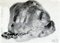
Fig. 163. External appearance of intact specimen. No. 1522. X0.75.
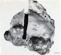
Fig. 164. External appearance of same specimen, showing where block was removed. X0.75.
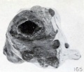
Fig. 165. Appearance of cross section of pregnant ovary and tube, same specimen. X0.75.
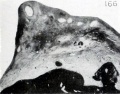
Fig. 166. Photograph of a section taken from the thick portion of the ovarian stroma near the mesovarium, showing a well-developed Graafian follicle. Same specimen. X2.25.
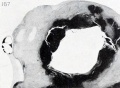
Fig. 167. Section of part of the same specimen, showing the clot which contains the empty vesicle largely surrounded by ovarian stroma. XI. 9.
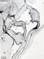
Fig. 168. Marked clubbing of villi of No. 550. X2.25.
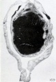
Fig. 169. Cross section of decidua and conceptus. No. 698. (See Chapter XII.) X4.5.
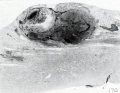
Fig. 170. Section of conceptus, decidua, and muscularis. No. 970. (See Chapter XII.) X4.5.
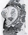
Fig. 171. Section of decidua and conceptus. No. 962. (See Chapter XII.) X4.5.
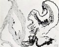
Fig. 172. Section of a part of the conceptus, showing chorionic membrane, cyemic (?) rudiment (x) and yolk-sac. No. 1843. (See Chapter XII.) X51.5.
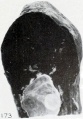
Fig. 173. External view of No. 2047, showing the distended amnion. (See Chapter XII.) X2.25.
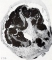
Fig. 174. Section of the tube and the conceptus. No. 977. (See Chapter XII.) X3.3.


| Embryology - 19 Apr 2024 |
|---|
| Google Translate - select your language from the list shown below (this will open a new external page) |
|
العربية | català | 中文 | 中國傳統的 | français | Deutsche | עִברִית | हिंदी | bahasa Indonesia | italiano | 日本語 | 한국어 | မြန်မာ | Pilipino | Polskie | português | ਪੰਜਾਬੀ ਦੇ | Română | русский | Español | Swahili | Svensk | ไทย | Türkçe | اردو | ייִדיש | Tiếng Việt These external translations are automated and may not be accurate. (More? About Translations) |
Mall FP. and Meyer AW. Studies on abortuses: a survey of pathologic ova in the Carnegie Embryological Collection. (1921) Contrib. Embryol., Carnegie Inst. Wash. Publ. 275, 12: 1-364.
- In this historic 1921 pathology paper, figures and plates of abnormal embryos are not suitable for young students.
1921 Carnegie Collection - Abnormal: Preface | 1 Collection origin | 2 Care and utilization | 3 Classification | 4 Pathologic analysis | 5 Size | 6 Sex incidence | 7 Localized anomalies | 8 Hydatiform uterine | 9 Hydatiform tubal | Chapter 10 Alleged superfetation | 11 Ovarian Pregnancy | 12 Lysis and resorption | 13 Postmortem intrauterine | 14 Hofbauer cells | 15 Villi | 16 Villous nodules | 17 Syphilitic changes | 18 Aspects | Bibliography | Figures | Contribution No.56 | Contributions Series | Embryology History
| Historic Disclaimer - information about historic embryology pages |
|---|
 |
File history
Click on a date/time to view the file as it appeared at that time.
| Date/Time | Thumbnail | Dimensions | User | Comment | |
|---|---|---|---|---|---|
| current | 19:12, 23 November 2012 |  | 940 × 1,200 (223 KB) | Z8600021 (talk | contribs) | ==Plate 16== Fig. 163. External appearance of intact specimen. No. 1522. X0.75. Fig. 164. External appearance of same specimen, showing where block was removed. X0.75. Fig. 165. Appearance of cross section of pregnant ovary and tube, same specimen. X0.7 |
You cannot overwrite this file.
File usage
The following 2 pages use this file: