File:Mall Meyer1921 plate11.jpg: Difference between revisions
(==Plate 11== Fig. 121. Slight epithelial proliferation on markedly hydatiform villi. No. 134. X50. Fig. 122. Extremely marked epithelial proliferation on a small hydatiform villus with glassy stroma. No. 415. X95. Fig. 123. The disintegrating capilla) |
No edit summary |
||
| Line 1: | Line 1: | ||
==Plate 11== | ==Plate 11== | ||
Fig. 121. Slight epithelial proliferation on markedly hydatiform villi. No. 134. X50. | Fig. 121. Slight epithelial proliferation on markedly hydatiform villi. No. 134. X50. | ||
| Line 14: | Line 12: | ||
Fig. 126. Villi showing only a trace of the capillaries. No. 651<7- X50. | Fig. 126. Villi showing only a trace of the capillaries. No. 651<7- X50. | ||
{{Mall Meyer1921Plate11}} | |||
{{Carnegie56 TOC}} | {{Carnegie56 TOC}} | ||
Latest revision as of 08:37, 6 December 2012
Plate 11
Fig. 121. Slight epithelial proliferation on markedly hydatiform villi. No. 134. X50.
Fig. 122. Extremely marked epithelial proliferation on a small hydatiform villus with glassy stroma. No. 415. X95.
Fig. 123. The disintegrating capillaries are represented merely by large, incomplete curved outlines. No. 749. X50.
Fig. 124. Collapsed capillaries in process of obliteration. No. 712. X95.
Fig. 125. Fenestrated, macerated, hydatiform villi among others which are quite fibrous. No. 651d. X50.
Fig. 126. Villi showing only a trace of the capillaries. No. 651<7- X50.
- Plate 11: Fig. 120 | Fig. 121 | Fig. 122 | Fig. 123 | Fig. 124 | Fig. 125 | Fig. 126 | Chapter 8. Hydatiform Degeneration in Uterine Pregnancy
| Embryology - 18 Apr 2024 |
|---|
| Google Translate - select your language from the list shown below (this will open a new external page) |
|
العربية | català | 中文 | 中國傳統的 | français | Deutsche | עִברִית | हिंदी | bahasa Indonesia | italiano | 日本語 | 한국어 | မြန်မာ | Pilipino | Polskie | português | ਪੰਜਾਬੀ ਦੇ | Română | русский | Español | Swahili | Svensk | ไทย | Türkçe | اردو | ייִדיש | Tiếng Việt These external translations are automated and may not be accurate. (More? About Translations) |
Mall FP. and Meyer AW. Studies on abortuses: a survey of pathologic ova in the Carnegie Embryological Collection. (1921) Contrib. Embryol., Carnegie Inst. Wash. Publ. 275, 12: 1-364.
- In this historic 1921 pathology paper, figures and plates of abnormal embryos are not suitable for young students.
1921 Carnegie Collection - Abnormal: Preface | 1 Collection origin | 2 Care and utilization | 3 Classification | 4 Pathologic analysis | 5 Size | 6 Sex incidence | 7 Localized anomalies | 8 Hydatiform uterine | 9 Hydatiform tubal | Chapter 10 Alleged superfetation | 11 Ovarian Pregnancy | 12 Lysis and resorption | 13 Postmortem intrauterine | 14 Hofbauer cells | 15 Villi | 16 Villous nodules | 17 Syphilitic changes | 18 Aspects | Bibliography | Figures | Contribution No.56 | Contributions Series | Embryology History
| Historic Disclaimer - information about historic embryology pages |
|---|
 |
File history
Click on a date/time to view the file as it appeared at that time.
| Date/Time | Thumbnail | Dimensions | User | Comment | |
|---|---|---|---|---|---|
| current | 19:45, 23 November 2012 | 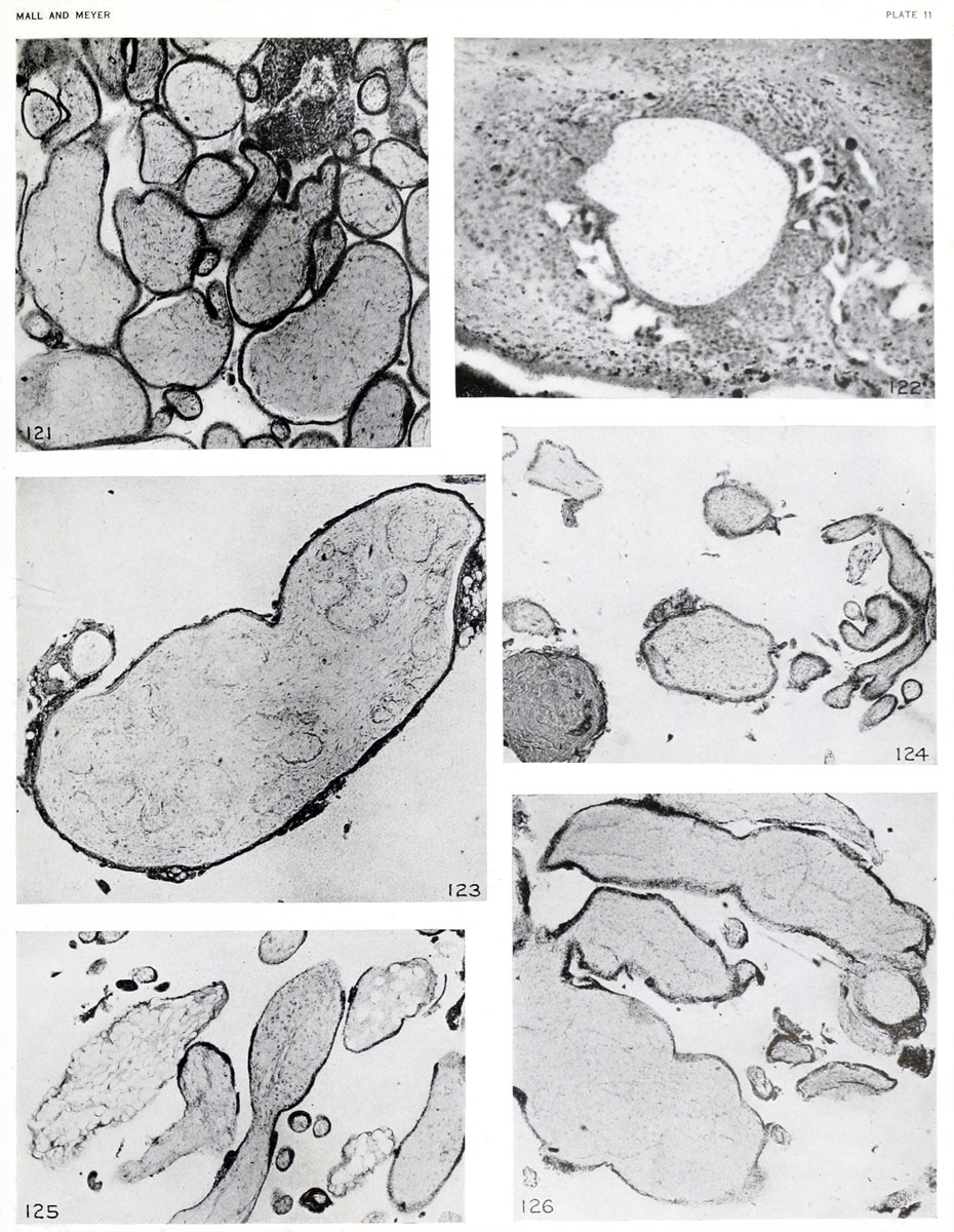 | 930 × 1,200 (272 KB) | Z8600021 (talk | contribs) | ==Plate 11== Fig. 121. Slight epithelial proliferation on markedly hydatiform villi. No. 134. X50. Fig. 122. Extremely marked epithelial proliferation on a small hydatiform villus with glassy stroma. No. 415. X95. Fig. 123. The disintegrating capilla |
You cannot overwrite this file.
File usage
The following 2 pages use this file: