File:Mall Meyer1921 fig218.jpg: Difference between revisions
No edit summary |
No edit summary |
||
| Line 2: | Line 2: | ||
No. 797. X1.35. | No. 797. X1.35. | ||
The abnormal curvatures, and especially the blunting of the extremities, are exemplified still better, and the drooping mandible, gaping mouth, and locally edematous cord are also well shown in No. 797, a fetus 35 mm. long, represented in figure 218. | |||
4) The specimen is covered with a few atrophic villi, and its wall is thin on one side and thick on the other. The interior is filled with a jelly-like magma and contains an atrophic embryo 35 mm., which is markedly deformed, having atrophic hands and feet and a very thin umbilical cord. The head is also reduced in size. | 4) The specimen is covered with a few atrophic villi, and its wall is thin on one side and thick on the other. The interior is filled with a jelly-like magma and contains an atrophic embryo 35 mm., which is markedly deformed, having atrophic hands and feet and a very thin umbilical cord. The head is also reduced in size. | ||
Latest revision as of 05:22, 4 December 2012
Fig. 218. A fetus and cord showing marked maceration changes
No. 797. X1.35.
The abnormal curvatures, and especially the blunting of the extremities, are exemplified still better, and the drooping mandible, gaping mouth, and locally edematous cord are also well shown in No. 797, a fetus 35 mm. long, represented in figure 218.
4) The specimen is covered with a few atrophic villi, and its wall is thin on one side and thick on the other. The interior is filled with a jelly-like magma and contains an atrophic embryo 35 mm., which is markedly deformed, having atrophic hands and feet and a very thin umbilical cord. The head is also reduced in size.
(5) Sections of the chorion show that it is hemorrhagic and inflamed. The chorionic wall is fibrous and degenerate, and the villi mostly necrotic, only a few of them being fairly preserved. They are bound together by fibrin and a great deal of fibrinoid substance. There is also considerable nuclear dust.
(6) Marked infiltration of the decidua; probably luetic.
- Plate 19: Figs. 217 | Figs. 218 | Fig. 219 | Fig. 220 | Fig. 221 | Fig. 222 | Fig. 223 | Fig. 224 | Fig. 225 | Fig. 226 | Fig. 227 | Fig. 228 | Fig. 229 | Fig. 230 | Fig. 231 | Fig. 232 | Fig. 233 | Fig. 234 | Chapter 13 Post-Mortem Intrauterine Changes
| Embryology - 19 Apr 2024 |
|---|
| Google Translate - select your language from the list shown below (this will open a new external page) |
|
العربية | català | 中文 | 中國傳統的 | français | Deutsche | עִברִית | हिंदी | bahasa Indonesia | italiano | 日本語 | 한국어 | မြန်မာ | Pilipino | Polskie | português | ਪੰਜਾਬੀ ਦੇ | Română | русский | Español | Swahili | Svensk | ไทย | Türkçe | اردو | ייִדיש | Tiếng Việt These external translations are automated and may not be accurate. (More? About Translations) |
Mall FP. and Meyer AW. Studies on abortuses: a survey of pathologic ova in the Carnegie Embryological Collection. (1921) Contrib. Embryol., Carnegie Inst. Wash. Publ. 275, 12: 1-364.
- In this historic 1921 pathology paper, figures and plates of abnormal embryos are not suitable for young students.
1921 Carnegie Collection - Abnormal: Preface | 1 Collection origin | 2 Care and utilization | 3 Classification | 4 Pathologic analysis | 5 Size | 6 Sex incidence | 7 Localized anomalies | 8 Hydatiform uterine | 9 Hydatiform tubal | Chapter 10 Alleged superfetation | 11 Ovarian Pregnancy | 12 Lysis and resorption | 13 Postmortem intrauterine | 14 Hofbauer cells | 15 Villi | 16 Villous nodules | 17 Syphilitic changes | 18 Aspects | Bibliography | Figures | Contribution No.56 | Contributions Series | Embryology History
| Historic Disclaimer - information about historic embryology pages |
|---|
 |
File history
Click on a date/time to view the file as it appeared at that time.
| Date/Time | Thumbnail | Dimensions | User | Comment | |
|---|---|---|---|---|---|
| current | 11:40, 3 December 2012 | 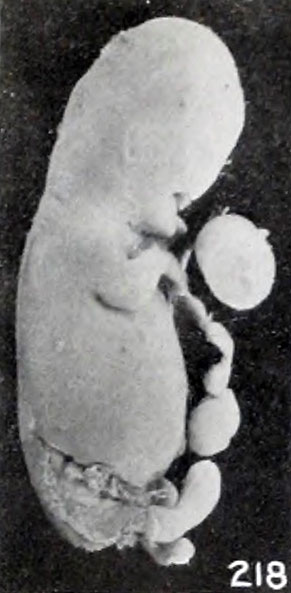 | 291 × 593 (31 KB) | Z8600021 (talk | contribs) | ==Fig. 218. A fetus and cord showing marked maceration changes== No. 797. X1.35. {{Mall Meyer1921Plate19}} {{Carnegie56 TOC}} |
You cannot overwrite this file.