File:Mall Meyer1921 fig19.jpg: Difference between revisions
(==Fig. 19. External appearance of chorion containing small nodular twin== No. 7886. Xl.15. :'''Plate 2''': Fig. 12 | Fig. 13 | [[:File:Mall_Meyer) |
No edit summary |
||
| Line 2: | Line 2: | ||
No. 7886. Xl.15. | No. 7886. Xl.15. | ||
One of the most interesting of these nodular embryos is No. 7886, received from Dr. Anfin Egdahl, shown in figure 19. This is one of double-ovum twins in which the chorionic vesicles were wholly distinct. The smaller of the twins is but 2 mm. long, with a greatest diameter of about the same dimension. The mate, on the contrary, is relatively a normally formed, somewhat stunted fetus, with a crown-rump length of 17 mm. The abortus containing the chorionic vesicle with the little nodule was somewhat larger than the one containing the fetus, although both chorionic vesicles were covered by approximately the same quantity of decidua. The dimensions of the chorionic vesicle belonging to the stunted embryo were 60 by 45 by 40 mm., and those of the one containing the nodular embryo 65 by 55 by 40 mm. The greater size of the latter was due probably to the greater distension of the chorionic membranes, which are thinner and possess only poorly developed villi. The other vesicle, on the contrary, shows very definite placental development, although one of its dimensions actually was smaller than the corresponding dimension of the chorionic vesicle belonging to the small nodule. Both specimens evidently were very decidedly macerated when aborted. | |||
:[[:File:Mall Meyer1921 plate02.jpg|'''Plate 2''']]: [[:File:Mall_Meyer1921_fig12.jpg|Fig. 12]] | [[:File:Mall_Meyer1921_fig13.jpg|Fig. 13]] | [[:File:Mall_Meyer1921_fig14.jpg|Fig. 14]] | [[:File:Mall_Meyer1921_fig15.jpg|Fig. 15]] | [[:File:Mall_Meyer1921_fig16.jpg|Fig. 16]] | [[:File:Mall_Meyer1921_fig13.jpg|Fig. 17]] | [[:File:Mall_Meyer1921_fig12.jpg|Fig. 17]] | [[:File:Mall_Meyer1921_fig18.jpg|Fig. 18]] | [[:File:Mall_Meyer1921_fig19.jpg|Fig. 19]] | [[Book - Contributions to Embryology Carnegie Institution No.56-4|Chapter 4 Pathologic analysis]] | :[[:File:Mall Meyer1921 plate02.jpg|'''Plate 2''']]: [[:File:Mall_Meyer1921_fig12.jpg|Fig. 12]] | [[:File:Mall_Meyer1921_fig13.jpg|Fig. 13]] | [[:File:Mall_Meyer1921_fig14.jpg|Fig. 14]] | [[:File:Mall_Meyer1921_fig15.jpg|Fig. 15]] | [[:File:Mall_Meyer1921_fig16.jpg|Fig. 16]] | [[:File:Mall_Meyer1921_fig13.jpg|Fig. 17]] | [[:File:Mall_Meyer1921_fig12.jpg|Fig. 17]] | [[:File:Mall_Meyer1921_fig18.jpg|Fig. 18]] | [[:File:Mall_Meyer1921_fig19.jpg|Fig. 19]] | [[Book - Contributions to Embryology Carnegie Institution No.56-4|Chapter 4 Pathologic analysis]] | ||
Revision as of 17:00, 24 November 2012
Fig. 19. External appearance of chorion containing small nodular twin
No. 7886. Xl.15.
One of the most interesting of these nodular embryos is No. 7886, received from Dr. Anfin Egdahl, shown in figure 19. This is one of double-ovum twins in which the chorionic vesicles were wholly distinct. The smaller of the twins is but 2 mm. long, with a greatest diameter of about the same dimension. The mate, on the contrary, is relatively a normally formed, somewhat stunted fetus, with a crown-rump length of 17 mm. The abortus containing the chorionic vesicle with the little nodule was somewhat larger than the one containing the fetus, although both chorionic vesicles were covered by approximately the same quantity of decidua. The dimensions of the chorionic vesicle belonging to the stunted embryo were 60 by 45 by 40 mm., and those of the one containing the nodular embryo 65 by 55 by 40 mm. The greater size of the latter was due probably to the greater distension of the chorionic membranes, which are thinner and possess only poorly developed villi. The other vesicle, on the contrary, shows very definite placental development, although one of its dimensions actually was smaller than the corresponding dimension of the chorionic vesicle belonging to the small nodule. Both specimens evidently were very decidedly macerated when aborted.
File history
Click on a date/time to view the file as it appeared at that time.
| Date/Time | Thumbnail | Dimensions | User | Comment | |
|---|---|---|---|---|---|
| current | 14:30, 24 November 2012 | 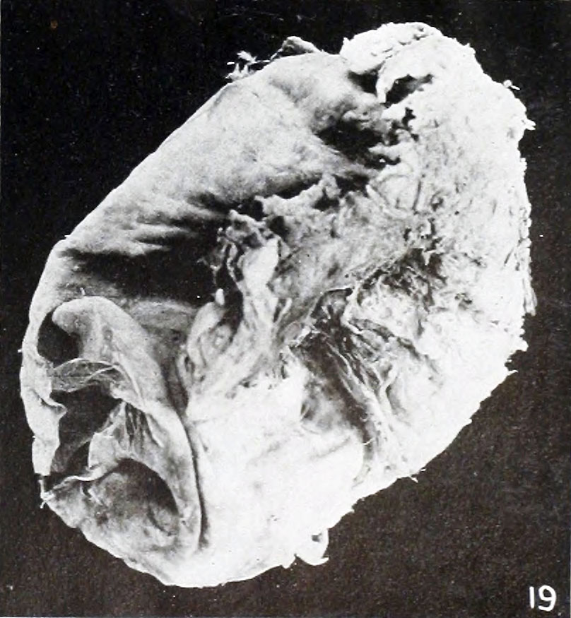 | 808 × 875 (134 KB) | Z8600021 (talk | contribs) | ==Fig. 19. External appearance of chorion containing small nodular twin== No. 7886. Xl.15. :'''Plate 2''': Fig. 12 | Fig. 13 | [[:File:Mall_Meyer |
You cannot overwrite this file.
File usage
The following 2 pages use this file: