File:Mall Meyer1921 fig18.jpg: Difference between revisions
(==Fig. 18. Cross-section of a nodular cyema, showing simple structure== No. 6510. X10. :'''Plate 2''': Fig. 12 | Fig. 13 | [[:File:Mall_Meyer1921) |
No edit summary |
||
| Line 3: | Line 3: | ||
No. 6510. X10. | No. 6510. X10. | ||
Although one would not suspect it from external appearances alone, this group of the pathologic is by far the most interesting. Some of these specimens show an astonishing simplicity in form and structure. Perhaps it was this fact which caused His (1891) to doubt whether they really were remnants of the whole embryo. His query is justified by the form and microscopic structure of many of them, as is so well illustrated by Nos. 2288, 1369, and 651#, shown in figures [[:File:Mall_Meyer1921_fig16.jpg|16]], [[:File:Mall_Meyer1921_fig17.jpg|17]], [[:File:Mall_Meyer1921_fig18.jpg|18]]. | |||
{{Mall_Meyer1921Plate2}} | |||
{{Carnegie56 TOC}} | |||
Latest revision as of 17:39, 24 November 2012
Fig. 18. Cross-section of a nodular cyema, showing simple structure
No. 6510. X10.
Although one would not suspect it from external appearances alone, this group of the pathologic is by far the most interesting. Some of these specimens show an astonishing simplicity in form and structure. Perhaps it was this fact which caused His (1891) to doubt whether they really were remnants of the whole embryo. His query is justified by the form and microscopic structure of many of them, as is so well illustrated by Nos. 2288, 1369, and 651#, shown in figures 16, 17, 18.
- Plate 2: Fig. 12 | Fig. 13 | Fig. 14 | Fig. 15 | Fig. 16 | Fig. 17 | Fig. 18 | Fig. 19 | Chapter 4 Pathologic analysis
| Embryology - 25 Apr 2024 |
|---|
| Google Translate - select your language from the list shown below (this will open a new external page) |
|
العربية | català | 中文 | 中國傳統的 | français | Deutsche | עִברִית | हिंदी | bahasa Indonesia | italiano | 日本語 | 한국어 | မြန်မာ | Pilipino | Polskie | português | ਪੰਜਾਬੀ ਦੇ | Română | русский | Español | Swahili | Svensk | ไทย | Türkçe | اردو | ייִדיש | Tiếng Việt These external translations are automated and may not be accurate. (More? About Translations) |
Mall FP. and Meyer AW. Studies on abortuses: a survey of pathologic ova in the Carnegie Embryological Collection. (1921) Contrib. Embryol., Carnegie Inst. Wash. Publ. 275, 12: 1-364.
- In this historic 1921 pathology paper, figures and plates of abnormal embryos are not suitable for young students.
1921 Carnegie Collection - Abnormal: Preface | 1 Collection origin | 2 Care and utilization | 3 Classification | 4 Pathologic analysis | 5 Size | 6 Sex incidence | 7 Localized anomalies | 8 Hydatiform uterine | 9 Hydatiform tubal | Chapter 10 Alleged superfetation | 11 Ovarian Pregnancy | 12 Lysis and resorption | 13 Postmortem intrauterine | 14 Hofbauer cells | 15 Villi | 16 Villous nodules | 17 Syphilitic changes | 18 Aspects | Bibliography | Figures | Contribution No.56 | Contributions Series | Embryology History
| Historic Disclaimer - information about historic embryology pages |
|---|
 |
File history
Click on a date/time to view the file as it appeared at that time.
| Date/Time | Thumbnail | Dimensions | User | Comment | |
|---|---|---|---|---|---|
| current | 14:30, 24 November 2012 | 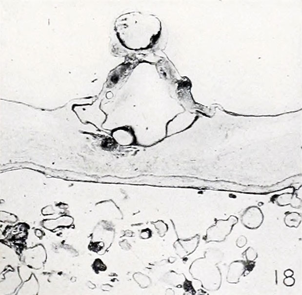 | 619 × 605 (62 KB) | Z8600021 (talk | contribs) | ==Fig. 18. Cross-section of a nodular cyema, showing simple structure== No. 6510. X10. :'''Plate 2''': Fig. 12 | Fig. 13 | [[:File:Mall_Meyer1921 |
You cannot overwrite this file.
File usage
The following 2 pages use this file: