File:Mall Meyer1921 fig166.jpg
Mall_Meyer1921_fig166.jpg (605 × 473 pixels, file size: 54 KB, MIME type: image/jpeg)
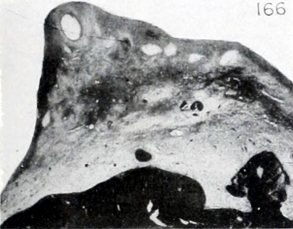
Fig. 166. Photograph of section from ovarian stroma near the mesovarium showing a Graafian follicle
Photograph of a section taken from the thick portion of the ovarian stroma near the mesovarium, showing a well-developed Graafian follicle. Same specimen. No. 1522. X2.25.
- Plate 16: Figs. 163 | Fig. 164 | Fig. 165 | Fig. 166 | Fig. 167 | Fig. 168 | Fig. 169 | Fig. 170 | Fig. 171 | Fig. 172 | Fig. 173 | Fig. 174 | Chapter 11 Ovarian Pregnancy
| Embryology - 23 Apr 2024 |
|---|
| Google Translate - select your language from the list shown below (this will open a new external page) |
|
العربية | català | 中文 | 中國傳統的 | français | Deutsche | עִברִית | हिंदी | bahasa Indonesia | italiano | 日本語 | 한국어 | မြန်မာ | Pilipino | Polskie | português | ਪੰਜਾਬੀ ਦੇ | Română | русский | Español | Swahili | Svensk | ไทย | Türkçe | اردو | ייִדיש | Tiếng Việt These external translations are automated and may not be accurate. (More? About Translations) |
Mall FP. and Meyer AW. Studies on abortuses: a survey of pathologic ova in the Carnegie Embryological Collection. (1921) Contrib. Embryol., Carnegie Inst. Wash. Publ. 275, 12: 1-364.
- In this historic 1921 pathology paper, figures and plates of abnormal embryos are not suitable for young students.
1921 Carnegie Collection - Abnormal: Preface | 1 Collection origin | 2 Care and utilization | 3 Classification | 4 Pathologic analysis | 5 Size | 6 Sex incidence | 7 Localized anomalies | 8 Hydatiform uterine | 9 Hydatiform tubal | Chapter 10 Alleged superfetation | 11 Ovarian Pregnancy | 12 Lysis and resorption | 13 Postmortem intrauterine | 14 Hofbauer cells | 15 Villi | 16 Villous nodules | 17 Syphilitic changes | 18 Aspects | Bibliography | Figures | Contribution No.56 | Contributions Series | Embryology History
| Historic Disclaimer - information about historic embryology pages |
|---|
 |
File history
Click on a date/time to view the file as it appeared at that time.
| Date/Time | Thumbnail | Dimensions | User | Comment | |
|---|---|---|---|---|---|
| current | 08:43, 13 December 2012 | | 605 × 473 (54 KB) | Z8600021 (talk | contribs) | ==Fig. 166. Photograph of section from ovarian stroma near the mesovarium showing a Graafian follicle== Photograph of a section taken from the thick portion of the ovarian stroma near the mesovarium, showing a well-developed Graafian follicle. Same spe |
You cannot overwrite this file.