File:Mall Meyer1921 fig14.jpg: Difference between revisions
(==Fig. 14. A fine hydatiform villous tree in section== No. 367. X 10. :'''Plate 2''': Fig. 12 | Fig. 13 | [[:File:Mall_Meyer1921_fig14.jpg|Fig.) |
No edit summary |
||
| Line 3: | Line 3: | ||
No. 367. X 10. | No. 367. X 10. | ||
As shown in section in [[:File:Mall_Meyer1921_fig14.jpg|figure 14]], some exceedingly fine hydatiform villous trees were found among the specimens in this group. Scaffoldings or frameworks, formed by the proliferating sycytium arising from the epithelium of the chorionic membrane, were also seen. Since syncytial buds were found far out on proliferations of trophoblast which capped the villi, and also in the center of trophoblastic nodules, the origin of the syncytium from the Langhans layer would seem to be exceptionally well illustrated. In some cases a detached hydatiform villus was fastened to two portions of the tube-wall. It is well to remember, however, that these attachments may have been gained, and indeed probably were gained, before the separation of the particular villus from the chorionic vesicle. | |||
{{Mall_Meyer1921Plate2}} | |||
{{Carnegie56 TOC}} | |||
Latest revision as of 17:27, 24 November 2012
Fig. 14. A fine hydatiform villous tree in section
No. 367. X 10.
As shown in section in figure 14, some exceedingly fine hydatiform villous trees were found among the specimens in this group. Scaffoldings or frameworks, formed by the proliferating sycytium arising from the epithelium of the chorionic membrane, were also seen. Since syncytial buds were found far out on proliferations of trophoblast which capped the villi, and also in the center of trophoblastic nodules, the origin of the syncytium from the Langhans layer would seem to be exceptionally well illustrated. In some cases a detached hydatiform villus was fastened to two portions of the tube-wall. It is well to remember, however, that these attachments may have been gained, and indeed probably were gained, before the separation of the particular villus from the chorionic vesicle.
- Plate 2: Fig. 12 | Fig. 13 | Fig. 14 | Fig. 15 | Fig. 16 | Fig. 17 | Fig. 18 | Fig. 19 | Chapter 4 Pathologic analysis
| Embryology - 20 Apr 2024 |
|---|
| Google Translate - select your language from the list shown below (this will open a new external page) |
|
العربية | català | 中文 | 中國傳統的 | français | Deutsche | עִברִית | हिंदी | bahasa Indonesia | italiano | 日本語 | 한국어 | မြန်မာ | Pilipino | Polskie | português | ਪੰਜਾਬੀ ਦੇ | Română | русский | Español | Swahili | Svensk | ไทย | Türkçe | اردو | ייִדיש | Tiếng Việt These external translations are automated and may not be accurate. (More? About Translations) |
Mall FP. and Meyer AW. Studies on abortuses: a survey of pathologic ova in the Carnegie Embryological Collection. (1921) Contrib. Embryol., Carnegie Inst. Wash. Publ. 275, 12: 1-364.
- In this historic 1921 pathology paper, figures and plates of abnormal embryos are not suitable for young students.
1921 Carnegie Collection - Abnormal: Preface | 1 Collection origin | 2 Care and utilization | 3 Classification | 4 Pathologic analysis | 5 Size | 6 Sex incidence | 7 Localized anomalies | 8 Hydatiform uterine | 9 Hydatiform tubal | Chapter 10 Alleged superfetation | 11 Ovarian Pregnancy | 12 Lysis and resorption | 13 Postmortem intrauterine | 14 Hofbauer cells | 15 Villi | 16 Villous nodules | 17 Syphilitic changes | 18 Aspects | Bibliography | Figures | Contribution No.56 | Contributions Series | Embryology History
| Historic Disclaimer - information about historic embryology pages |
|---|
 |
File history
Click on a date/time to view the file as it appeared at that time.
| Date/Time | Thumbnail | Dimensions | User | Comment | |
|---|---|---|---|---|---|
| current | 14:27, 24 November 2012 | 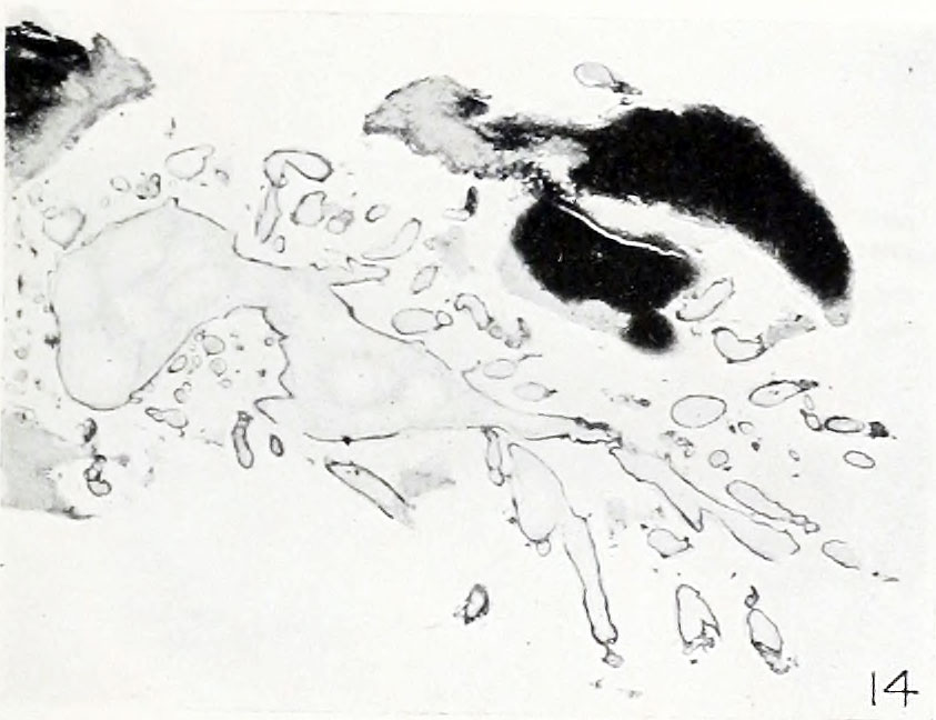 | 843 × 648 (69 KB) | Z8600021 (talk | contribs) | ==Fig. 14. A fine hydatiform villous tree in section== No. 367. X 10. :'''Plate 2''': Fig. 12 | Fig. 13 | [[:File:Mall_Meyer1921_fig14.jpg|Fig. |
You cannot overwrite this file.
File usage
The following 2 pages use this file: