File:Mall Meyer1921 fig09.jpg

Original file (1,009 × 711 pixels, file size: 134 KB, MIME type: image/jpeg)
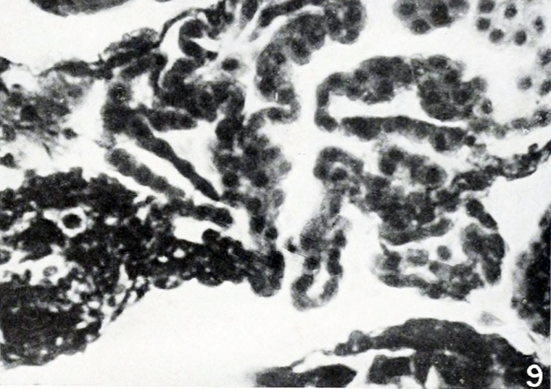
Fig. 9. Festoons of chorionic epithelium
No. 1324. X300.
Changes simulating those of lues were noticed in no tubal conceptuses in this group, but several excellent examples of hydatiform degeneration were found in Nos. 415, 602, 686, 772, and 889. According to Seitz (19040, the occurrence of hydatiform moles was observed in connection with tubal pregnancies by Freund, Matwejew and Sykow, Otto, and Wenzel. Others no doubt have observed it since then, but as only a few villi are contained in a single cross-section of the tube, and but few cross-sections of each specimen were examined in our series, one can not be certain of one's diagnosis in every instance. If more villi were present this difficulty would be obviated, although it must be remembered that a large series of specimens necessarily supplement each other. Furthermore, the changes in many villi are so typical, both as to outward form and structure, as to be undoubted. Since many of the villi were decidedly degenerate, one could hardly expect to find much proliferation of the endothelium, but remarkable specimens, such as that in figure 9, were occasionally found. In some cases the presence of hydatiform degeneration became probable only through comparison of the villi in question with those found in many undoubted cases of hydatiform degeneration examined previously.
| Embryology - 19 Apr 2024 |
|---|
| Google Translate - select your language from the list shown below (this will open a new external page) |
|
العربية | català | 中文 | 中國傳統的 | français | Deutsche | עִברִית | हिंदी | bahasa Indonesia | italiano | 日本語 | 한국어 | မြန်မာ | Pilipino | Polskie | português | ਪੰਜਾਬੀ ਦੇ | Română | русский | Español | Swahili | Svensk | ไทย | Türkçe | اردو | ייִדיש | Tiếng Việt These external translations are automated and may not be accurate. (More? About Translations) |
Mall FP. and Meyer AW. Studies on abortuses: a survey of pathologic ova in the Carnegie Embryological Collection. (1921) Contrib. Embryol., Carnegie Inst. Wash. Publ. 275, 12: 1-364.
- In this historic 1921 pathology paper, figures and plates of abnormal embryos are not suitable for young students.
1921 Carnegie Collection - Abnormal: Preface | 1 Collection origin | 2 Care and utilization | 3 Classification | 4 Pathologic analysis | 5 Size | 6 Sex incidence | 7 Localized anomalies | 8 Hydatiform uterine | 9 Hydatiform tubal | Chapter 10 Alleged superfetation | 11 Ovarian Pregnancy | 12 Lysis and resorption | 13 Postmortem intrauterine | 14 Hofbauer cells | 15 Villi | 16 Villous nodules | 17 Syphilitic changes | 18 Aspects | Bibliography | Figures | Contribution No.56 | Contributions Series | Embryology History
| Historic Disclaimer - information about historic embryology pages |
|---|
 |
File history
Click on a date/time to view the file as it appeared at that time.
| Date/Time | Thumbnail | Dimensions | User | Comment | |
|---|---|---|---|---|---|
| current | 13:50, 24 November 2012 | | 1,009 × 711 (134 KB) | Z8600021 (talk | contribs) | ==Fig. 9. Festoons of chorionic epithelium== No. 1324. X300. :'''Plate 1''': Fig 6 | Fig 7 | Fig 8 | [[:Mall_Meyer1921_fig09. |
You cannot overwrite this file.
File usage
The following 2 pages use this file: