File:Mall1912-fig11.jpg: Difference between revisions
From Embryology
(Z8600021 uploaded a new version of File:Mall1912-fig11.jpg) |
mNo edit summary |
||
| (2 intermediate revisions by the same user not shown) | |||
| Line 1: | Line 1: | ||
==Fig. 11. View from below of a model of the heart of an embryo 11 mm long== | ==Fig. 11. View from below of a model of the heart of an embryo 11 mm long== | ||
(No. | (No. {{CE353}}). X 50. The ventricles have been cut off. The connective tissue septa are colored yellow. | ||
* the right cushion marks the center of the anlage of the anterior and posterior cusps of the tricuspid valve, and the left the anlage of the posterior cusp of the mitral valve. The septum aorto pulmonale soon blends with the cushions through a dorso—lateral wing which is divided into two branches to encircle in part the right Venous ostium, one of which blends with the right lateral endocardial thickening, and ‘the other, thepmedial, blends with the anterior process of the medial valves now represented by the right lower wing of the anterior endocardial cushion. It is thus seen that through the blending of the septum aorto pulmonale with the right side of the anterior endocardial cushion, the right venous ostium is nearly encircled by endothelial connective tissue. This connection may still be seen in the adult heart where the septum aorto-pulmonale (the tendon of the conus) is found to blend with the fibrous ring of the right ostium at the anterior border of the attachment of the medial cusp of the tricuspid valve. | |||
{{Mall1912 figures}} | {{Mall1912 figures}} | ||
[[Category:Carnegie Embryo 353]] | |||
Latest revision as of 17:27, 31 July 2017
Fig. 11. View from below of a model of the heart of an embryo 11 mm long
(No. 353). X 50. The ventricles have been cut off. The connective tissue septa are colored yellow.
- the right cushion marks the center of the anlage of the anterior and posterior cusps of the tricuspid valve, and the left the anlage of the posterior cusp of the mitral valve. The septum aorto pulmonale soon blends with the cushions through a dorso—lateral wing which is divided into two branches to encircle in part the right Venous ostium, one of which blends with the right lateral endocardial thickening, and ‘the other, thepmedial, blends with the anterior process of the medial valves now represented by the right lower wing of the anterior endocardial cushion. It is thus seen that through the blending of the septum aorto pulmonale with the right side of the anterior endocardial cushion, the right venous ostium is nearly encircled by endothelial connective tissue. This connection may still be seen in the adult heart where the septum aorto-pulmonale (the tendon of the conus) is found to blend with the fibrous ring of the right ostium at the anterior border of the attachment of the medial cusp of the tricuspid valve.
| Mall Figure Legend | ||
|---|---|---|
|
|
|
| Historic Disclaimer - information about historic embryology pages |
|---|
 |
- Links: Fig 1 | Fig 2 | Fig 3 | Fig 4 | Fig 5 | Fig 6 | Fig 7 | Fig 8 | Fig 9 | Fig 10 | Fig 11 | Fig 12 | Fig 13 | Fig 14 | Fig 15 | Fig 16 | Fig 17 | Fig 18 | Fig 19 | Fig 20 | Fig 21 | Fig 22 | Fig 23 | Fig 24-25 | Fig 26 | Fig 27 | Fig 28 | Fig 31-33 | Mall 1912 | Historic Papers | Carnegie Collection | Franklin Mall | Cardiovascular Development

Reference
Mall FP. On the development of the human heart. (1912) Amer. J Anat. 13: 249-298.
Cite this page: Hill, M.A. (2024, April 19) Embryology Mall1912-fig11.jpg. Retrieved from https://embryology.med.unsw.edu.au/embryology/index.php/File:Mall1912-fig11.jpg
- © Dr Mark Hill 2024, UNSW Embryology ISBN: 978 0 7334 2609 4 - UNSW CRICOS Provider Code No. 00098G
File history
Click on a date/time to view the file as it appeared at that time.
| Date/Time | Thumbnail | Dimensions | User | Comment | |
|---|---|---|---|---|---|
| current | 16:43, 29 October 2015 | 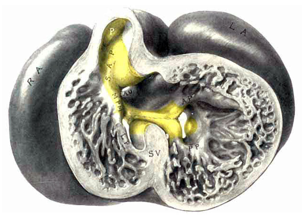 | 1,000 × 725 (124 KB) | Z8600021 (talk | contribs) | |
| 16:42, 29 October 2015 | 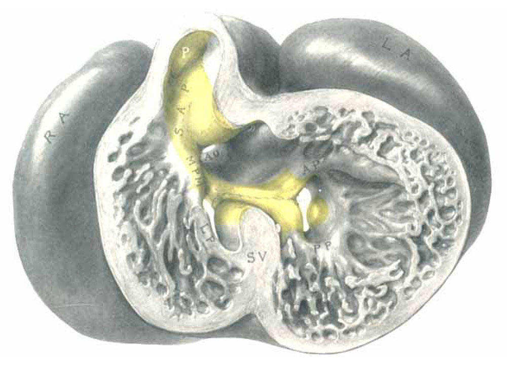 | 1,000 × 725 (98 KB) | Z8600021 (talk | contribs) | ||
| 14:56, 29 October 2015 | 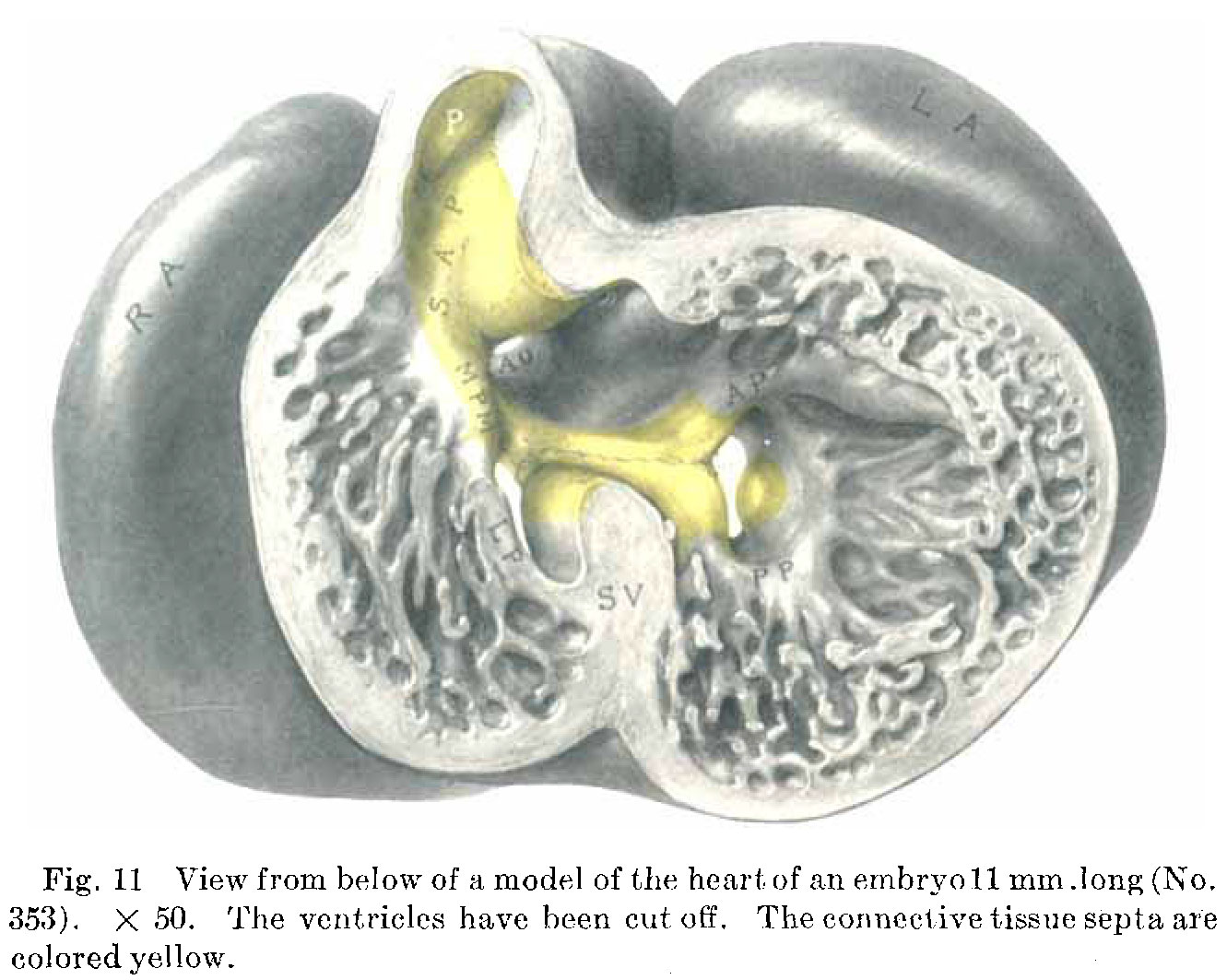 | 1,331 × 1,063 (202 KB) | Z8600021 (talk | contribs) | {{Mall1912 figures}} |
You cannot overwrite this file.
File usage
The following page uses this file: