File:Mall1906-fig08.jpg
From Embryology

Size of this preview: 578 × 599 pixels. Other resolution: 726 × 753 pixels.
Original file (726 × 753 pixels, file size: 75 KB, MIME type: image/jpeg)
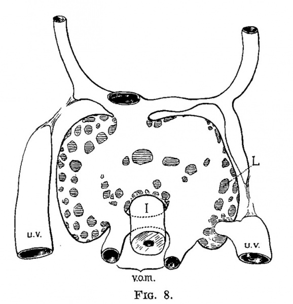
Fig. 8. Semi-diagrammatic reconstruction of the veins of the liver of a human embryo 4.3 mm long
Carnegie Embryo (No. 148)
| At this time the umbilical veins are broken in their course, having already passed the first stage of their growth, and from now on are destined to pass through the liver rather than past it (fig. 8). It is interesting to note that the primary sinusoidal liver—that portion which arises with the omphalo-mesenteric vein—is formed while the umbilical vein empties directly into the ductus Cuvieri. The process is at its height in an embryo of about the same age (No. 76), as shown in fig. 9 and fig. 10. The growth of the liver around and through the omphalo-mesenteric veins is accompanied by the growth of capillaries from this vein into the new anlage. Hand in hand with this process the circulation through the omphalo-mesenteric veins is further reduced by the growth and enlargement of the umbilical veins. |
|
| Historic Disclaimer - information about historic embryology pages |
|---|
| Pages where the terms "Historic" (textbooks, papers, people, recommendations) appear on this site, and sections within pages where this disclaimer appears, indicate that the content and scientific understanding are specific to the time of publication. This means that while some scientific descriptions are still accurate, the terminology and interpretation of the developmental mechanisms reflect the understanding at the time of original publication and those of the preceding periods, these terms, interpretations and recommendations may not reflect our current scientific understanding. (More? Embryology History | Historic Embryology Papers) |
- Links: Fig 6 | Fig 7 | Fig 8 | Fig 9 | Fig 10 | | Fig 11 | Fig 12 | Fig 13 | Fig 14 | Fig 15 | Fig 16 | Fig 17 | Fig 18 | Fig 19 | Fig 20 | Fig 21 | Fig 22 | Fig 23 | Fig 24 | Fig 25 | Fig 26 | Fig 27 | Fig 28 | Fig 29 | Fig 30 | Mall 1906 | Historic Embryology Papers | Liver Development
Reference
Mall FP. A study of the structural unit of the liver. (1906) Amer. J Anat. 5:227-308.
Cite this page: Hill, M.A. (2024, April 16) Embryology Mall1906-fig08.jpg. Retrieved from https://embryology.med.unsw.edu.au/embryology/index.php/File:Mall1906-fig08.jpg
- © Dr Mark Hill 2024, UNSW Embryology ISBN: 978 0 7334 2609 4 - UNSW CRICOS Provider Code No. 00098G
File history
Click on a date/time to view the file as it appeared at that time.
| Date/Time | Thumbnail | Dimensions | User | Comment | |
|---|---|---|---|---|---|
| current | 19:00, 20 September 2015 |  | 726 × 753 (75 KB) | Z8600021 (talk | contribs) | |
| 19:00, 20 September 2015 |  | 1,328 × 1,154 (325 KB) | Z8600021 (talk | contribs) | {{Mall1906liverfigures}} |
You cannot overwrite this file.
File usage
The following 3 pages use this file:
