File:Mall1891 Plate02Fig01.jpg
From Embryology

Size of this preview: 449 × 599 pixels. Other resolution: 623 × 831 pixels.
Original file (623 × 831 pixels, file size: 125 KB, MIME type: image/jpeg)
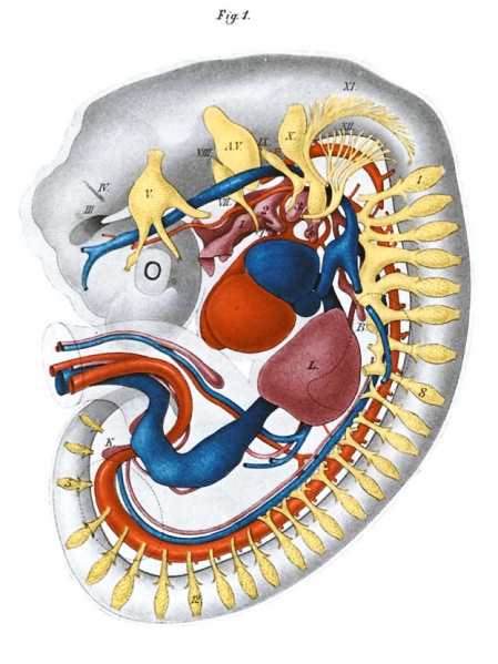
Plate XXX Figure 1. Reconstruction viewed from the left side
Enlarged 15 times.
The dotted lines mark the extremities.
Legend
- HI., IV., V., etc. - cranial nerves
- A. V. - auditory vesicle
- I. 2, 3, and 4 - branchial pockets
- T. - thyroid gland
- B. - bronchus
- L. - liver
- K. - kidney;
- yellow - nerves
- red - arteries
- blue - veins
| Week: | 1 | 2 | 3 | 4 | 5 | 6 | 7 | 8 |
| Carnegie stage: | 1 2 3 4 | 5 6 | 7 8 9 | 10 11 12 13 | 14 15 | 16 17 | 18 19 | 20 21 22 23 |
| Historic Disclaimer - information about historic embryology pages |
|---|
 |
- Links: Fig 1 Reconstruction viewed from the left side | Fig 2 Gastric diverticulum | Plate 1 Fig 1 External view | Plate 1 Fig 2 Pericardial and pleuro-peritoneal cavities | Plate 2 Fig 1 Reconstruction viewed from the left side | Plate 2 Fig 2 Reconstruction viewed from the left side | Carnegie stage 15
Reference
Mall FP. A human embryo twenty-six days old. (1891) J Morphol. 5: 459-480.
Cite this page: Hill, M.A. (2024, April 18) Embryology Mall1891 Plate02Fig01.jpg. Retrieved from https://embryology.med.unsw.edu.au/embryology/index.php/File:Mall1891_Plate02Fig01.jpg
- © Dr Mark Hill 2024, UNSW Embryology ISBN: 978 0 7334 2609 4 - UNSW CRICOS Provider Code No. 00098G
File history
Click on a date/time to view the file as it appeared at that time.
| Date/Time | Thumbnail | Dimensions | User | Comment | |
|---|---|---|---|---|---|
| current | 12:15, 11 March 2012 | | 623 × 831 (125 KB) | Z8600021 (talk | contribs) | ==Mall 1891 Plate 2 Fig01== Category:Franklin Mall |
You cannot overwrite this file.