File:Magnetic Resonance Imaging in an adult patient with tetralogy of Fallot.jpg: Difference between revisions
(The MRI shows a) Shows Dilation and hypertrophy of right ventricle b) Shows the stenotic and tortuous homograft conduit responsible for the dilated and hypertrophied right ventricle. © 2009 Bailliard and Anderson; licensee BioMed Central Ltd. This is ) |
No edit summary |
||
| Line 2: | Line 2: | ||
a) Shows Dilation and hypertrophy of right ventricle | a) Shows Dilation and hypertrophy of right ventricle | ||
b) Shows the stenotic and tortuous homograft conduit responsible for the dilated and hypertrophied right ventricle. | b) Shows the stenotic and tortuous homograft conduit responsible for the dilated and hypertrophied right ventricle. | ||
© 2009 Bailliard and Anderson; licensee BioMed Central Ltd. | © 2009 Bailliard and Anderson; licensee BioMed Central Ltd. | ||
This is an Open Access article distributed under the terms of the Creative Commons Attribution License (http://creativecommons.org/licenses/by/2.0), | This is an Open Access article distributed under the terms of the Creative Commons Attribution License (http://creativecommons.org/licenses/by/2.0), which permits unrestricted use, distribution, and reproduction in any medium, provided the original work is properly cited. | ||
which permits unrestricted use, distribution, and reproduction in any medium, provided the original work is properly cited. | |||
PMID: 19144126 | PMID: 19144126 | ||
Journal URL: http://www.ncbi.nlm.nih.gov/pmc/articles/PMC2651859/?tool=pubmed | Journal URL: http://www.ncbi.nlm.nih.gov/pmc/articles/PMC2651859/?tool=pubmed | ||
Image URL: http://www.ncbi.nlm.nih.gov/pmc/articles/PMC2651859/figure/F9/ | Image URL: http://www.ncbi.nlm.nih.gov/pmc/articles/PMC2651859/figure/F9/ | ||
{{Template:2011 Student Image}} | {{Template:2011 Student Image}} | ||
Latest revision as of 23:06, 5 October 2011
The MRI shows
a) Shows Dilation and hypertrophy of right ventricle
b) Shows the stenotic and tortuous homograft conduit responsible for the dilated and hypertrophied right ventricle.
© 2009 Bailliard and Anderson; licensee BioMed Central Ltd. This is an Open Access article distributed under the terms of the Creative Commons Attribution License (http://creativecommons.org/licenses/by/2.0), which permits unrestricted use, distribution, and reproduction in any medium, provided the original work is properly cited.
PMID: 19144126
Journal URL: http://www.ncbi.nlm.nih.gov/pmc/articles/PMC2651859/?tool=pubmed Image URL: http://www.ncbi.nlm.nih.gov/pmc/articles/PMC2651859/figure/F9/
- Note - This image was originally uploaded as part of a student project and may contain inaccuracies in either description or acknowledgements. Students have been advised in writing concerning the reuse of content and may accidentally have misunderstood the original terms of use. If image reuse on this non-commercial educational site infringes your existing copyright, please contact the site editor for immediate removal.
Cite this page: Hill, M.A. (2024, April 20) Embryology Magnetic Resonance Imaging in an adult patient with tetralogy of Fallot.jpg. Retrieved from https://embryology.med.unsw.edu.au/embryology/index.php/File:Magnetic_Resonance_Imaging_in_an_adult_patient_with_tetralogy_of_Fallot.jpg
- © Dr Mark Hill 2024, UNSW Embryology ISBN: 978 0 7334 2609 4 - UNSW CRICOS Provider Code No. 00098G
File history
Click on a date/time to view the file as it appeared at that time.
| Date/Time | Thumbnail | Dimensions | User | Comment | |
|---|---|---|---|---|---|
| current | 23:04, 5 October 2011 | 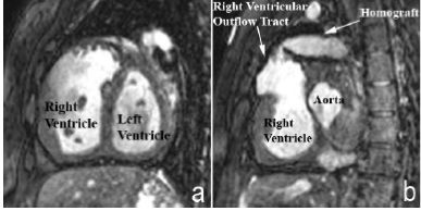 | 387 × 194 (17 KB) | Z3291423 (talk | contribs) | The MRI shows a) Shows Dilation and hypertrophy of right ventricle b) Shows the stenotic and tortuous homograft conduit responsible for the dilated and hypertrophied right ventricle. © 2009 Bailliard and Anderson; licensee BioMed Central Ltd. This is |
You cannot overwrite this file.
File usage
There are no pages that use this file.