File:Macklin1914 plate13.jpg
From Embryology
Size of this preview: 556 × 599 pixels. Other resolution: 2,226 × 2,399 pixels.
Original file (2,226 × 2,399 pixels, file size: 575 KB, MIME type: image/jpeg)
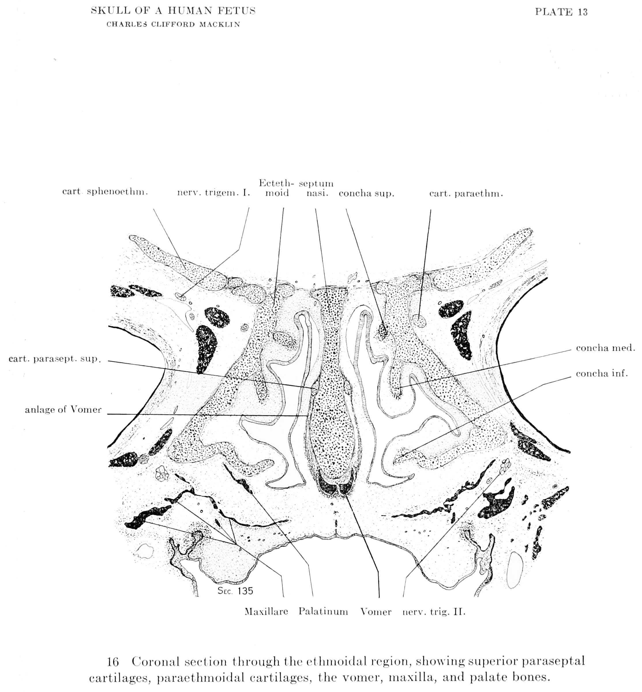
Plate 13.
Fig. 16. Coronal section through the ethinoidal region, showing superior paraseptal cartilages, paraethnioidal cartilages, the vomer, maxilla, and palate bones.
| Historic Disclaimer - information about historic embryology pages |
|---|
 |
- Links: plate 1 | plate 2 | plate 3 | plate 4 | plate 5 | plate 6 | fig 6 | fig 7 | plate 7 | fig 8 | fig 9 | plate 8 | fig 10 | plate 9 | fig 11 | fig 12 | plate 10 | plate 11 | fig 14 | Macklin 1914 part 1 | Macklin 1914 part 2 | Skull Development
Reference
Macklin CC. The skull of a human fetus of 40 mm 1. (1914) Amer. J Anat. 16(3): 317-386.
Cite this page: Hill, M.A. (2024, April 18) Embryology Macklin1914 plate13.jpg. Retrieved from https://embryology.med.unsw.edu.au/embryology/index.php/File:Macklin1914_plate13.jpg
- © Dr Mark Hill 2024, UNSW Embryology ISBN: 978 0 7334 2609 4 - UNSW CRICOS Provider Code No. 00098G
File history
Click on a date/time to view the file as it appeared at that time.
| Date/Time | Thumbnail | Dimensions | User | Comment | |
|---|---|---|---|---|---|
| current | 20:53, 26 June 2016 | | 2,226 × 2,399 (575 KB) | Z8600021 (talk | contribs) | ==Plate 13.== Fig. 15. Coronal section through the dorsal part of ethmoidal region, showing posterior cupidar process, the nasal septum, the interorbital sejitum with attached alae, and the palatine and vomer. {{Macklin1914 figures}} |
You cannot overwrite this file.
File usage
The following page uses this file: