File:Macklin1914 plate02.jpg

Original file (2,196 × 2,608 pixels, file size: 746 KB, MIME type: image/jpeg)
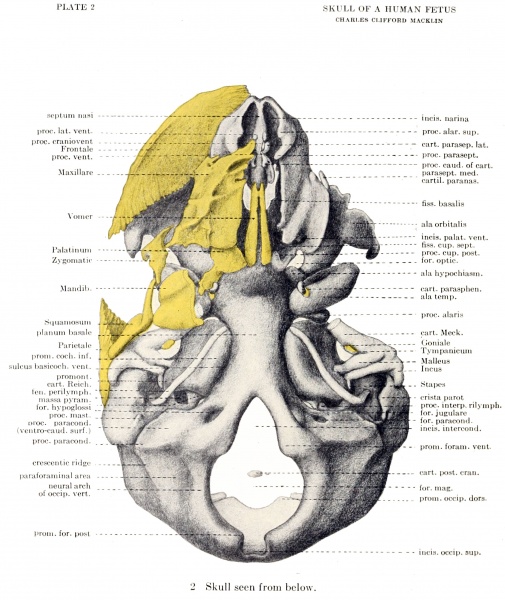
Plate 2. Wax plate reconstruction of the chondrocranium of a 40mm human fetus
Seen from below.
Regarded from below (fig. 2) the model shows in the foreground the mandibular (not shown in fig. 2) and upper part of the hyoid arches. Behind these we see the semi-cylindrical ventral surface of the planum basale, separated from the elongated, flattened, ovoid partes cochleares by deep furrows. It is to be observed that the anterior extremities of the latter do not project beyond the planum. The forked structure at the root of the planum, perforated for the hypoglossal nerve, is seen to be the anterior commencement of the flattened, ring-like sides of the foramen magnum, a downwardly projecting angle marking the position of the future occipital condyle. Lateral to and above the condyle there appears a stout cartilaginous process, which supports the lower and anterior part of the otic capsule. This is known as the processus paracondyloideus (Voit), and above it is seen a wide opening, the jugular foramen (fig. 4).
The two Meckelian cartilages (fig. 3) enclose an angle, rather sharp ventrally, in which are found the structures of the floor of the mouth. The inwardly curved palatine bones, with the assistance of the inner laminae of the pterygoid processes (fig. 2), imperfectly cut off the posterior part of the nasal cavity, and between the pterygoid laminae and the planum basale is seen the space occupied by the naso-pharynx (fig. 10) . Attached to these medial laminae, and indeed developed from their caudal tips, are the cartilagines parasphenoidales (Voit, '09), the representatives of the later hamular processes. The pterygoid laminae are quite separate from the alae temporales, which appear, from this viewpoint, as rhomboidal, perforated blocks.
Lying along the caudal border of the nasal septum are seen the anterior paraseptal cartilages in front, and the long thin plates of the vomer behind (fig. 2); and within each nasal cavity is a small mass of cartilage, lying in the middle meatus (fig. 12), to which the name oariilago meatus medii has been given and whose significance will be discussed with the regio ethmoidalis.
| Historic Disclaimer - information about historic embryology pages |
|---|
 |
- Links: plate 1 | plate 2 | plate 3 | plate 4 | plate 5 | plate 6 | fig 6 | fig 7 | plate 7 | fig 8 | fig 9 | plate 8 | fig 10 | plate 9 | fig 11 | fig 12 | plate 10 | plate 11 | fig 14 | Macklin 1914 part 1 | Macklin 1914 part 2 | Skull Development
Reference
Macklin CC. The skull of a human fetus of 40 mm 1. (1914) Amer. J Anat. 16(3): 317-386.
Cite this page: Hill, M.A. (2024, April 18) Embryology Macklin1914 plate02.jpg. Retrieved from https://embryology.med.unsw.edu.au/embryology/index.php/File:Macklin1914_plate02.jpg
- © Dr Mark Hill 2024, UNSW Embryology ISBN: 978 0 7334 2609 4 - UNSW CRICOS Provider Code No. 00098G
File history
Click on a date/time to view the file as it appeared at that time.
| Date/Time | Thumbnail | Dimensions | User | Comment | |
|---|---|---|---|---|---|
| current | 22:04, 20 June 2016 | | 2,196 × 2,608 (746 KB) | Z8600021 (talk | contribs) |
You cannot overwrite this file.
File usage
The following 2 pages use this file: