File:Macklin-plate04.jpg
Original file (2,331 × 3,061 pixels, file size: 956 KB, MIME type: image/jpeg)
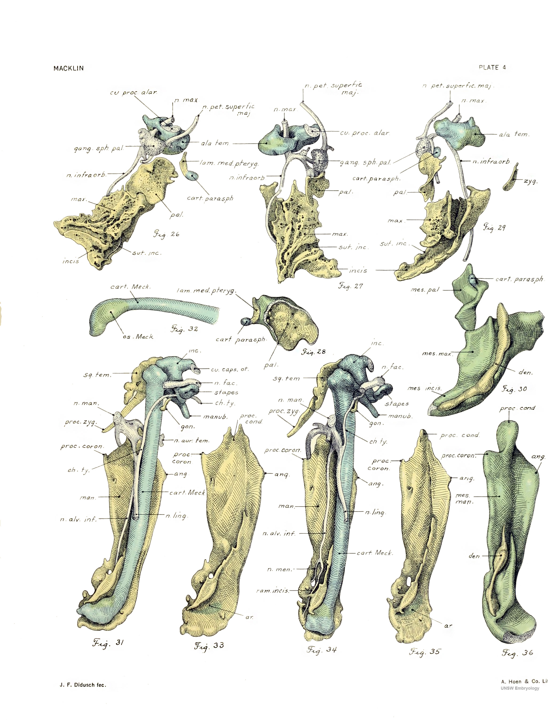
Plate 4. The skull of a human fetus of 43 millimeters greatest length
By Charles C. Macklin. (5 plates containing 47 figures)
All drawings were made by Mr. James F. Didusch according to geometric projection. With the exception of figure 7, which was made from a profile reconstruction, all figures were drawn from the original plaster-of-paris models made from human fetus No. 886 of the collection of the Carnegie Laboratory of Embryology. The number of the model from which each figure was drawn is given, together with the magnification.
Note - the magnifications refer to the original print versions, not the online images.
- Links: Plate 1 | Plate 2 | Plate 3 | Plate 4 | No.48 Early Fetal Skull
| Historic Disclaimer - information about historic embryology pages |
|---|
 |
Fig. 26. Right maxilla, palate, medial pterygoid plate with hamular process, temporal wing, sphenopalatine ganglion, and associated nerves
Right maxilla, palate, medial pterygoid plate with hamular process, temporal wing, sphenopalatine ganglion, and associated nerves, seen directly from within. Cut surface showing junction of temporal wing with alar process is seen. Note the incisive suture partially separating the maxilla and premaxilla. Model 11. X12.5.
Fig. 27. Right maxilla, palate, temporal wing, sphenopalatine ganglion, and associated nerves
The same structures as those seen in figure 26 with the exception of the medial pterygoid plate, seen from above. The relations of the maxillary division of the trigeminal nerve and its branches to the different structures are shown. Model 11. X12.5.
Fig. 28. Lateral aspect of the right palate bone and medial pterygoid plate
Lateral aspect of the right palate bone and medial pterygoid plate, with their investment of condensed mesenchyme. Model 18. X12.5.
Fig. 29. Lateral aspect of the right palate bone and medial pterygoid plate
The same structures seen in figures 26 and 27, but viewed from below. The tooth gutter of the maxilla and premaxilla is conspicuous and the lower end of the incisive suture appears. The zygomatic bone also is shown. Compare with figure 30. Model 11. X12.5.
Fig. 30. Condensed mesenchyme enveloping the right maxilla, palate, and medial pterygoid plate
The developing teeth are seen in their gutter in the maxilla. (Compare with figure 29 drawn from approximately the same point of view.) The cartilaginous hamular process is seen projecting from the medial pterygoid lamina. Compare also with figure 36, forbdeveloping teeth of the right lower jaw. Model 25. X12.5.
Fig. 31. Frank view of the right mandible, Meckel's cartilage
Frank view of the right mandible, Meckel's cartilage, and associated structures, seen from within. The cartilaginous precursors of the auditory ossicles are seen above, in relation to the facial and chorda tympani nerves. A ghmpse of the squama temporalis is given and also of the goniale. Note the relations of the mandibular division of the trigeminal nerve. Model 9. X12.5.
Fig. 32. Lateral aspect of lower end of the right Meckel's cartilage
Lateral aspect of lower end of the right Meckel's cartilage, showing especially the area applied closely to the mandible, where the cartilage is showing the changes preliminary to ossification. (Compare with figures 33 and 35.) Model 16. X12.5.
Fig. 33. Right mandible
Right mandible from same viewpoint as in figure 31, showing the tooth gutter and area of close apposition to the lower end of Meckel's cartilage. Model 10. X12.5.
Fig. 34. Right mandible
The same structures as those shown in figure 31, but the model was rotated somewhat medially around its long axis. While presenting all the structures from a new angle, it shows especially the relation of the nerves to the mandible. Model 9. X12.5.
Fig. 35. View of right mandible with the model
View of right mandible with the model in the same position as that shown in figure 34; it is rotated so as to afford a good view of the tooth gutter. Model 10. X12.5.
Fig. 36. Condensed mesenchyme around right mandible
Condensed mesenchyme around right mandible viewed from approximately the same point as that of figure 35. It shows the developing teeth of the right lower jaw. The mesenchyme is connected across the midhne with its partner of the opposite side, the cut edge being shown. Model 23. X12.5.
| Historic Disclaimer - information about historic embryology pages |
|---|
|
Glossary Links
- Glossary: A | B | C | D | E | F | G | H | I | J | K | L | M | N | O | P | Q | R | S | T | U | V | W | X | Y | Z | Numbers | Symbols | Term Link
Cite this page: Hill, M.A. (2024, April 19) Embryology Macklin-plate04.jpg. Retrieved from https://embryology.med.unsw.edu.au/embryology/index.php/File:Macklin-plate04.jpg
- © Dr Mark Hill 2024, UNSW Embryology ISBN: 978 0 7334 2609 4 - UNSW CRICOS Provider Code No. 00098G
File history
Click on a date/time to view the file as it appeared at that time.
| Date/Time | Thumbnail | Dimensions | User | Comment | |
|---|---|---|---|---|---|
| current | 15:53, 23 April 2014 | | 2,331 × 3,061 (956 KB) | Z8600021 (talk | contribs) | |
| 10:41, 16 February 2011 | 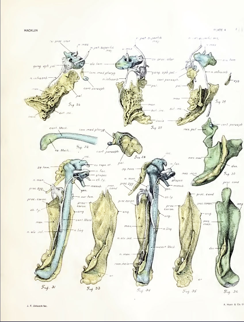 | 848 × 1,115 (192 KB) | S8600021 (talk | contribs) | ==Plate 4. The skull of a human fetus of 43 millimeters greatest length== By Charles C. Macklin. (5 plates containing 47 figures) All drawings were made by Mr. James F. Didusch according to geometric projection. With the exception of figure 7, which was |
You cannot overwrite this file.
File usage
The following 3 pages use this file: