File:Macklin-plate02.jpg: Difference between revisions
mNo edit summary |
mNo edit summary |
||
| Line 1: | Line 1: | ||
==Plate 2. The skull of a human fetus of 43 millimeters greatest length== | ==Plate 2. The skull of a human fetus of 43 millimeters greatest length== | ||
{{Macklin1921 figures}} | {| | ||
| {{Macklin1921 figures}} | |||
In general, blue is used to indicate cartilage and precartilage, yellow to indicate bone, and green for beginning ossification centers. Cut edges are white. The chief departures from this scheme are as follows: | In general, blue is used to indicate cartilage and precartilage, yellow to indicate bone, and green for beginning ossification centers. Cut edges are white. The chief departures from this scheme are as follows: | ||
Plate 2. — Figure 7, brain green; figure 8, mesenchyme green; figure 11, mucous membrane of pharynx yellow; figures 12 and 13, paraseptal cartilage and precartilage green. | Plate 2. — Figure 7, brain green; figure 8, mesenchyme green; figure 11, mucous membrane of pharynx yellow; figures 12 and 13, paraseptal cartilage and precartilage green. | ||
| valign=bottom| | |||
===Virtual Slide=== | |||
{{SlideMacklin1921Plate2}} | |||
|} | |||
===Fig. 5. Skull from right side, showing membrane bones=== | ===Fig. 5. Skull from right side, showing membrane bones=== | ||
Latest revision as of 13:53, 24 April 2014
Plate 2. The skull of a human fetus of 43 millimeters greatest length
All drawings were made by Mr. James F. Didusch according to geometric projection. With the exception of figure 7, which was made from a profile reconstruction, all figures were drawn from the original plaster-of-paris models made from human fetus No. 886 of the collection of the Carnegie Laboratory of Embryology. The number of the model from which each figure was drawn is given, together with the magnification. Note - the magnifications refer to the original print versions, not the online images. ReferenceMacklin CC. the skull of a human fetus of 43 millimeters greatest length. (1921) Contrib. Embryol., Carnegie Inst. Wash. Publ., 48, 10:59-102. Cite this page: Hill, M.A. (2024, April 16) Embryology Macklin-plate02.jpg. Retrieved from https://embryology.med.unsw.edu.au/embryology/index.php/File:Macklin-plate02.jpg
In general, blue is used to indicate cartilage and precartilage, yellow to indicate bone, and green for beginning ossification centers. Cut edges are white. The chief departures from this scheme are as follows: Plate 2. — Figure 7, brain green; figure 8, mesenchyme green; figure 11, mucous membrane of pharynx yellow; figures 12 and 13, paraseptal cartilage and precartilage green. |
Virtual Slide
|
Fig. 5. Skull from right side, showing membrane bones
The cervical vertebrae and cartilaginous branchial arch skeleton are included. Only the right half of the skull is shown. Model 1. X6.25.
Fig. 6. Left half of chondrocranium
Left half of chondrocranium, cervical vertebrae, and cartilaginous branchial arch skeleton as seen from left side. Model 1. X6.25.
Fig. 7. Profiles of external form of head
Profiles of external form of head, brain and upper end of spinal cord, and skull, in their normal relation to one another, as seen from the right side. Drawn from a profile reconstruction. Xl-9.
Fig. 8. Condensed mesenchyme enveloping the vomer
Condensed mesenchyme enveloping the vomer, seen from front, side, and above. The anterior extremities of the vomer are indicated. The gutter in the center is for the lower edge of the nasal septum. There is a slight amount of lateral curvature. The cut edges of the mesenchyme are indicated. Model 22. X12.5.
Fig. 9. Two halves of the vomer
Two halves of the vomer from the same point of view as that of figure 8. They are very slender spicules of bone lying along the lower border of the nasal septum. Model 21. X12.5.
Fig. 10. Median stem of skull as seen from right side
Median stem of skull as seen from right side It consists of the basal plate behind and the interorbital and nasal septa in front, forming ai- "btuse angle at the body of the sphenoid. The adjoining exoccipital cartilage is shown in part. Junctions with cartilage lying laterally are shown. Model 4. X12.5.
Fig. 11. Right half of basal plate and parts of upper two cervical vertebrae
Right half of basal plate and parts of upper two cervical vertebrae, sectioned in the mid-sagittal plane. The cut surface is seen in frank view. Shows the preossification center for the basioccipital, the notochord, the pharyngeal bursa with a little of the epithelium of the roof of the pharynx, the temporal wing, dorsum seUae and a portion of the exoccipital. Model 8. X12.5.
Fig. 12. Left cartilage of Jacobson
Left cartilage of Jacobson from left side with neighboring septum. Models 2 and 25. Xl2,5.
Fig. 13. Cartilages of Jacobson
Cartilages of Jacobson from below in relation to nasal septum. Models 2 and 25. X12.5.
Fig. 14. Right half of occipital cartilage and parietal plate
Right half of occipital cartilage and parietal plate from in front and within, with the ascending process and right half of inter- parietal bone in its mesenchyme. Connections with adjoining cartilages are shown. Model 24. X6.25.
| Historic Disclaimer - information about historic embryology pages |
|---|
 |
Glossary Links
- Glossary: A | B | C | D | E | F | G | H | I | J | K | L | M | N | O | P | Q | R | S | T | U | V | W | X | Y | Z | Numbers | Symbols | Term Link
Cite this page: Hill, M.A. (2024, April 16) Embryology Macklin-plate02.jpg. Retrieved from https://embryology.med.unsw.edu.au/embryology/index.php/File:Macklin-plate02.jpg
- © Dr Mark Hill 2024, UNSW Embryology ISBN: 978 0 7334 2609 4 - UNSW CRICOS Provider Code No. 00098G
File history
Click on a date/time to view the file as it appeared at that time.
| Date/Time | Thumbnail | Dimensions | User | Comment | |
|---|---|---|---|---|---|
| current | 16:27, 23 April 2014 | 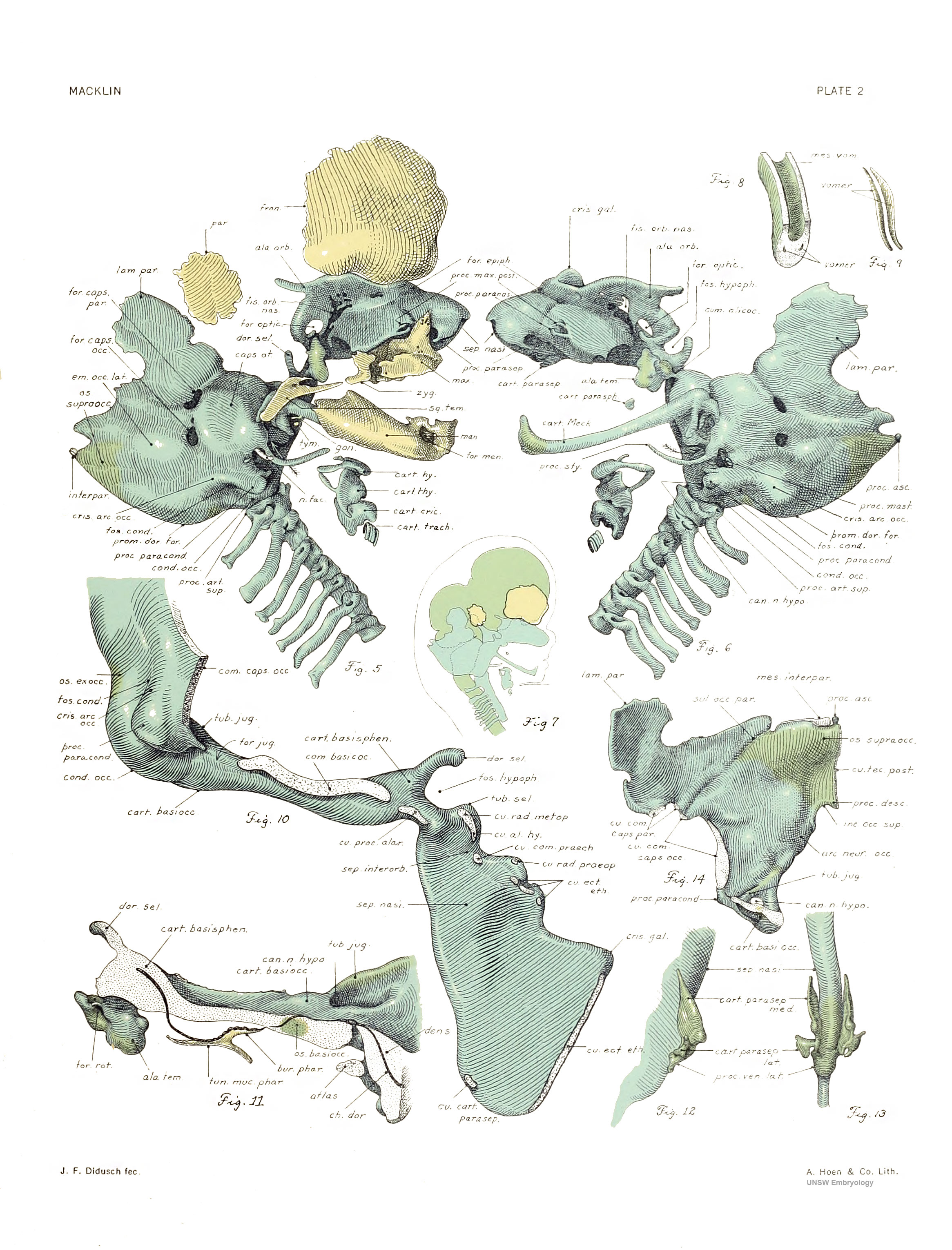 | 2,331 × 3,061 (1.08 MB) | Z8600021 (talk | contribs) | |
| 10:25, 16 February 2011 |  | 846 × 1,113 (196 KB) | S8600021 (talk | contribs) | ==Plate 2. The skull of a human fetus of 43 millimeters greatest length== By Charles C. Macklin. (5 plates containing 47 figures) All drawings were made by Mr. James F. Didusch according to geometric projection. With the exception of figure 7, which was |
You cannot overwrite this file.
File usage
The following 3 pages use this file: