File:MERS-CoV EM1.jpg
MERS-CoV_EM1.jpg (537 × 537 pixels, file size: 68 KB, MIME type: image/jpeg)
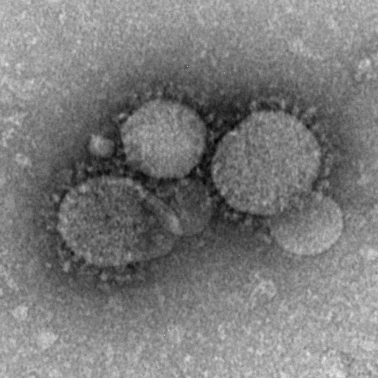
MERS-CoV particles as seen by negative stain electron microscopy, coronavirus virions contain characteristic "corona" of club-like projections from the viral membrane.
Reference
National Center for Immunization and Respiratory Diseases (NCIRD), Division of Viral Diseases
CDC Source: Cynthia Goldsmith/Maureen Metcalfe/Azaibi Tamin
Copyright
Information that is created by or for the US government is within the public domain. Public domain information on CDC Stacks may be freely distributed and copied. However, it is requested that in any subsequent use of this work, CDC be given appropriate acknowledgment.
Cite this page: Hill, M.A. (2024, April 20) Embryology MERS-CoV EM1.jpg. Retrieved from https://embryology.med.unsw.edu.au/embryology/index.php/File:MERS-CoV_EM1.jpg
- © Dr Mark Hill 2024, UNSW Embryology ISBN: 978 0 7334 2609 4 - UNSW CRICOS Provider Code No. 00098G
File history
Click on a date/time to view the file as it appeared at that time.
| Date/Time | Thumbnail | Dimensions | User | Comment | |
|---|---|---|---|---|---|
| current | 00:31, 21 January 2020 | | 537 × 537 (68 KB) | Z8600021 (talk | contribs) | MERS-CoV particles as seen by negative stain electron microscopy. Virions contain characteristic club-like projections emanating from the viral membrane. ===Reference=== CDC Source: Cynthia Goldsmith/Maureen Metcalfe/Azaibi Tamin {{Footer}} Category:VirusCategory:EM |
You cannot overwrite this file.
File usage
The following 3 pages use this file: