File:Low 07.jpg: Difference between revisions
No edit summary |
No edit summary |
||
| Line 1: | Line 1: | ||
==Fig. 7== | ==Fig. 7 Model of the Pharynx== | ||
Seen from the left side and slightly in front. | |||
The mouth is represented as a narrow transverse fissure between the forebrain and the body wall. In its roof is a slight funnel-shaped out-pushing - the commencement of the pocket of ectoderm to form the hypophysis. The mouth is separated from the pharynx by a complete buccopharyngeal membrane. The pharynx widens out rapidly behind this, and is flattened dorso-ventrally (fig.7). | |||
There are four pairs of pharyngeal pouches, of which the third and fourth are small and pointed. The second pocket is so situated that it lies much more ventral than caudal to the first pocket. The first two pairs of pockets are elongated vertically, and come in direct contact with the external ectoderm, where it forms the bottoms of the first and second pairs of visceral clefts (fig.7). | |||
{{Low 1908}} | {{Low 1908}} | ||
Latest revision as of 02:37, 22 February 2012
Fig. 7 Model of the Pharynx
Seen from the left side and slightly in front.
The mouth is represented as a narrow transverse fissure between the forebrain and the body wall. In its roof is a slight funnel-shaped out-pushing - the commencement of the pocket of ectoderm to form the hypophysis. The mouth is separated from the pharynx by a complete buccopharyngeal membrane. The pharynx widens out rapidly behind this, and is flattened dorso-ventrally (fig.7).
There are four pairs of pharyngeal pouches, of which the third and fourth are small and pointed. The second pocket is so situated that it lies much more ventral than caudal to the first pocket. The first two pairs of pockets are elongated vertically, and come in direct contact with the external ectoderm, where it forms the bottoms of the first and second pairs of visceral clefts (fig.7).
A 13-14 somite stage embryo would be similar to a Carnegie stage 11 (23 - 26 days) Somite Number 13 - 20.
- 13-14 Somite Paper: Plate 1 | Plate 2 | Plate 3 | Fig 1 | Fig 2 | Fig 3 | Fig 4 | Fig 5 | Fig 6 | Fig 7 | Fig 8 | Fig 9 | Fig 10 | Fig 11 | Fig 12 | Fig 13 | Fig 14 | Fig 15
| Historic Disclaimer - information about historic embryology pages |
|---|
 |
Reference
Low A. Description of a human embryo of 13-14 mesodermic somites. (1908) J Anat Physiol. 42(3): 237-51. PMID 17232769 | PMC1289161
Cite this page: Hill, M.A. (2024, April 19) Embryology Low 07.jpg. Retrieved from https://embryology.med.unsw.edu.au/embryology/index.php/File:Low_07.jpg
- © Dr Mark Hill 2024, UNSW Embryology ISBN: 978 0 7334 2609 4 - UNSW CRICOS Provider Code No. 00098G
File history
Click on a date/time to view the file as it appeared at that time.
| Date/Time | Thumbnail | Dimensions | User | Comment | |
|---|---|---|---|---|---|
| current | 23:46, 20 February 2012 | 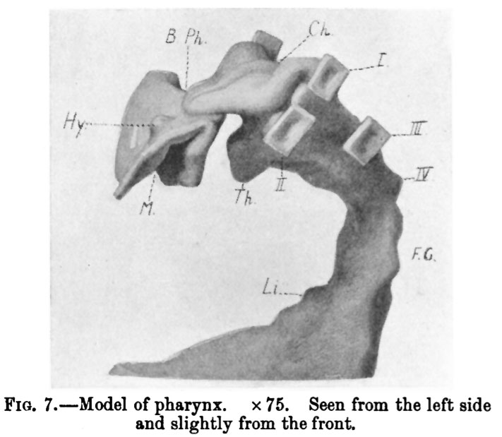 | 711 × 619 (56 KB) | S8600021 (talk | contribs) |
You cannot overwrite this file.
File usage
The following 2 pages use this file: