File:Locy1895 plate26.jpg: Difference between revisions
(==Plate XXVL== The ligures are from untouched negatives and are noteworthy in showing the very early condition of the optic vesicle, the accessory vesicles and in some cases the primitive rnetameric segrnenta They are all photographs of spart irre-tr...) |
(Z8600021 uploaded a new version of File:Locy1895 plate26.jpg) |
(No difference)
| |
Revision as of 15:17, 27 August 2018
Plate XXVL
The ligures are from untouched negatives and are noteworthy in showing the very early condition of the optic vesicle, the accessory vesicles and in some cases the primitive rnetameric segrnenta They are all photographs of spart irre-träfe and, with the exception of Fig. J, are X about 20 diameters.
Fig. 1. PhotograPh of embryo between Balfour’s stages i? and c.
FIg. 2. somewhat older embryo. The embryonic rim on the left side shows faintly Some of the metameric segments (not reproduced by the artist).
Fig. 3. Slightly older embryo to show the forrnation of the heachplate and the Central wedgeshaped Process thereon. This is the stage in which the neural folds are started along the margins of the body. They are ventrally curved.
Fig. 4. stage with a rounded head-Plate when the optic vesicles first become evident.
Fig. 5. Another embryo, about the same age as the Preceding, in which the optic vesicles and one of the accessory vesicles are shown.
Fig. 6. Somewhat older embryo showing the infolding for the optic vesicles extending across the median Plane.
Fig. 7. Older embryo somewhat higher magnifred The headsplate is very broad, the trunlc narrow. The neural folds of the head lie nearly in the horizontal Plane. The rnetarneric segments show well on the left margin of the head-P1ate. They exist in the earlier stages, but are very diiiicult to catch with the camera.
Fig. 8. speeirnen in which the dePressions for the optic vesicles show very distinctly
Figs. 9 and 10. Two ernbryos older (although smaller) than the preceding. In both two Pairs of accessory optic vesicles are to be seen on the cephalic Plate baclc of the Primary optic vesicles.
Figs. 11 and 12. Two specimens slightly older than the preceding two, seen from different Point of observation
Fig. 13. Embryo after the neural folds have begun to grow uPwards, seen obliquely from the left sicle. Gives an external view of— the vesicles on one side and an internal view of them on the oPPosite side of the neural folda
Fig. 14. Embryo from which Fig. 29, Plate XXV1I, is drawjx Shows metaIneric segments on the exPosed ventral surface of the neural folds.
FIg. 15. Embryo of same age as Fig. 13 and Fig. Hi, Plate XXVII. shows metarneric segments on the neural folds in front of the eye vesiclea
Fig. 16. Embryo of the same age as Fig. 9 just above it. Seen in a Position more favorable to bring out the accessory vesicles on the cephalic plate. The optic vesicle on the right side shows as an external Protuberance.
Fig. 17. somewhat older embryo viewed obliquely from above. shows the optic vesicle of the right side as an external rounded Protuberance, and that of the left side from within as a cup. Behind the optic vesiele on the left side of the cephalic place are four accessory vesiclea They show their serial reiation with the Primary optic vesicle.
FIg. 18. specimen showing a large development of the central tongueislilte Process. Embryo slightly older than that in Fig. 7. 586 Lock.
Figs. 19 and 20. Side view of two heads of embryos vvith open neural groove to show the external appearance of the optic vesicle
Fig. 21. Embryo of same age as Fig. 13 viewed obliquely from above.
Fig. 22. Side view of embryo of the same age (with broadly open neural gross-re) to show the external appearance of the optic vesicie and the other vesicles behind it.
Fig. 23. Embryo after partie-l closure of the neural groove Showing two openings, an anterior and a posterior one, into the neural canaL
Fig. 24. Some-what older specimen with head broken off. showing three openings (sma1ler than those in the preceding embryo) into the neural canaL
| Historic Disclaimer - information about historic embryology pages |
|---|
 |
Reference
Locy WA.Contribution to the structure and development of the vertebrate head. (1895) J. Morphol. 11(3): 497-595.
Cite this page: Hill, M.A. (2024, April 16) Embryology Locy1895 plate26.jpg. Retrieved from https://embryology.med.unsw.edu.au/embryology/index.php/File:Locy1895_plate26.jpg
- © Dr Mark Hill 2024, UNSW Embryology ISBN: 978 0 7334 2609 4 - UNSW CRICOS Provider Code No. 00098G
File history
Click on a date/time to view the file as it appeared at that time.
| Date/Time | Thumbnail | Dimensions | User | Comment | |
|---|---|---|---|---|---|
| current | 15:17, 27 August 2018 | 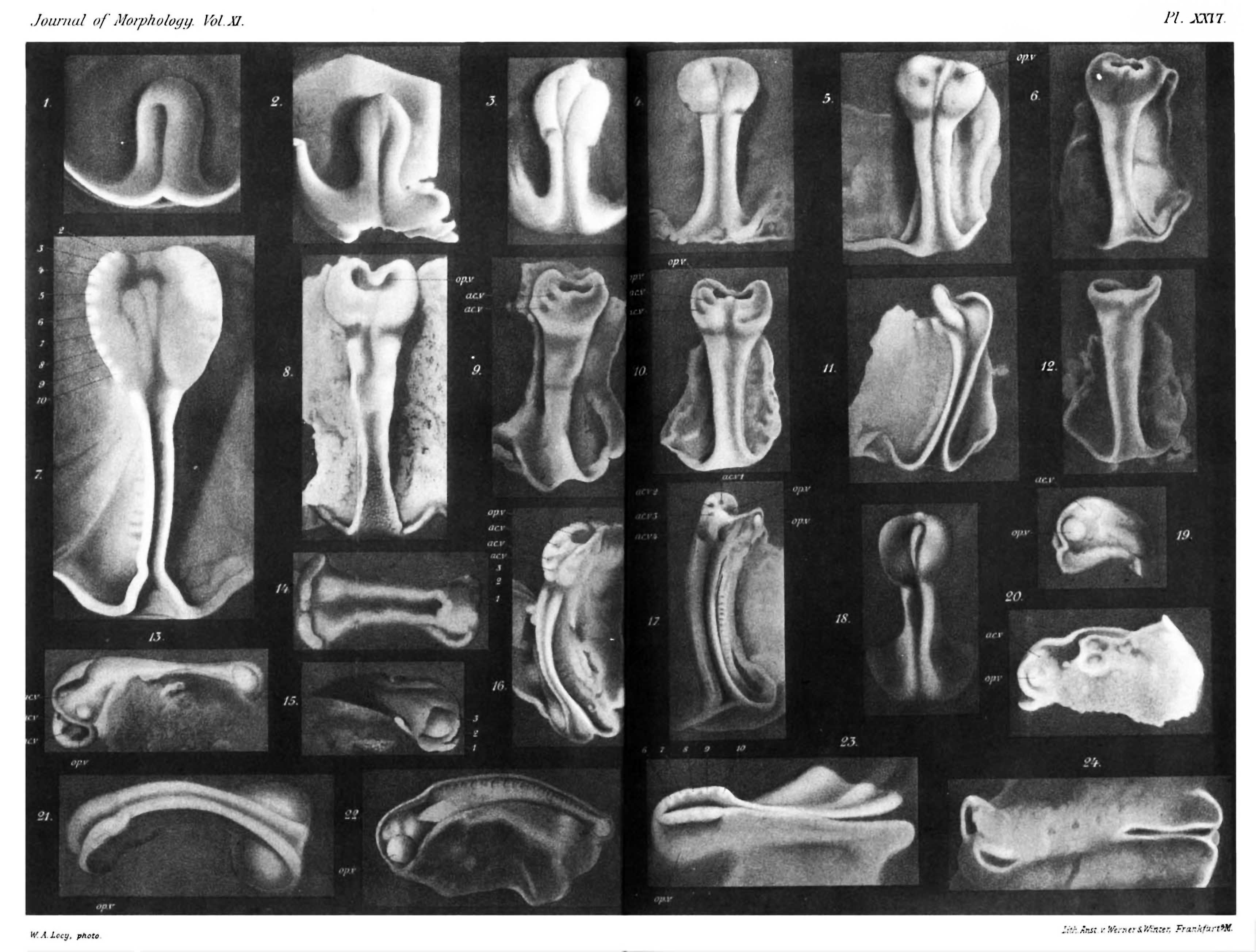 | 3,440 × 2,608 (439 KB) | Z8600021 (talk | contribs) | |
| 15:16, 27 August 2018 | 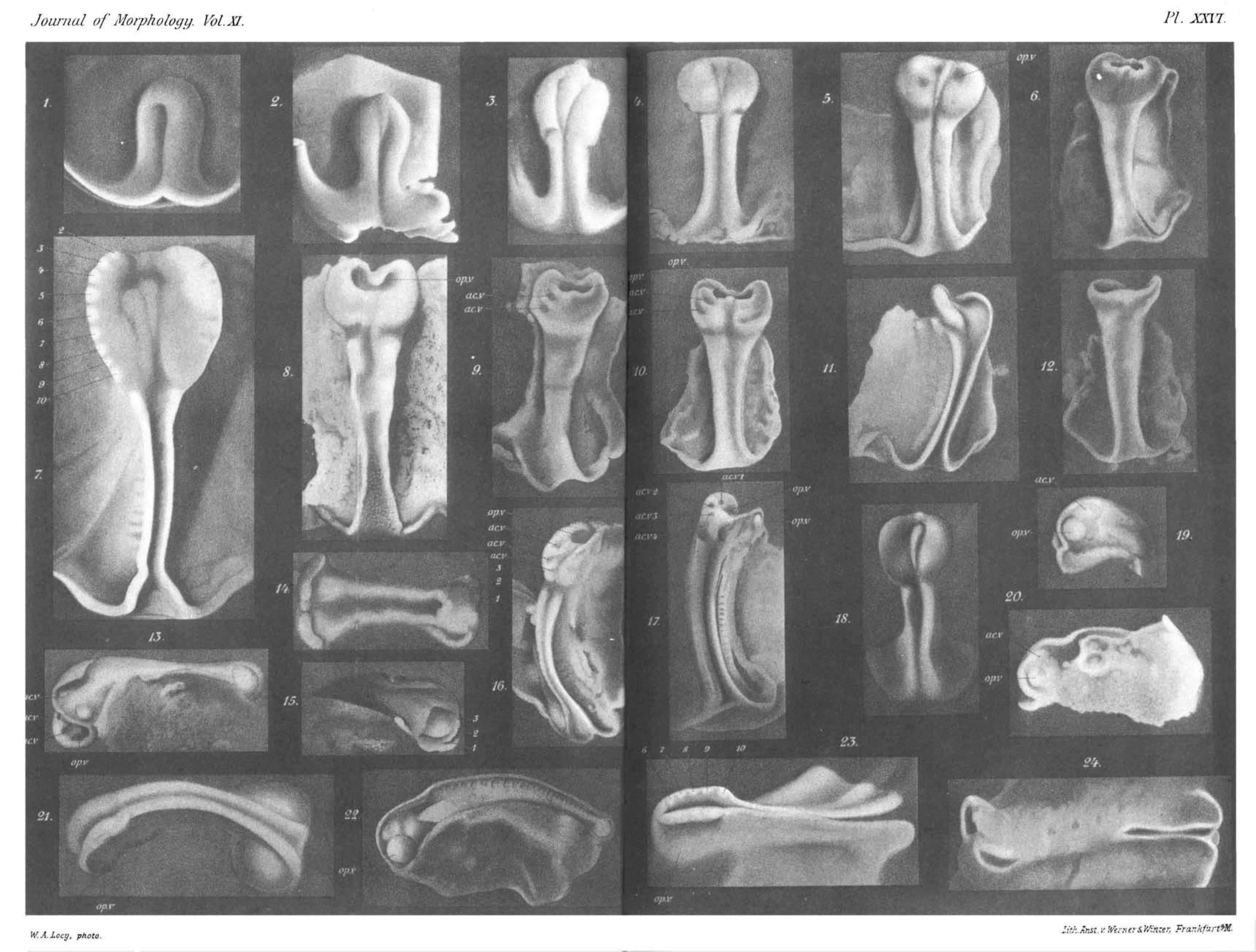 | 3,440 × 2,608 (417 KB) | Z8600021 (talk | contribs) | ==Plate XXVL== The ligures are from untouched negatives and are noteworthy in showing the very early condition of the optic vesicle, the accessory vesicles and in some cases the primitive rnetameric segrnenta They are all photographs of spart irre-tr... |
You cannot overwrite this file.
File usage
The following page uses this file: