File:Lockwood1887b fig41.jpg
From Embryology
No higher resolution available.
Lockwood1887b_fig41.jpg (800 × 547 pixels, file size: 129 KB, MIME type: image/jpeg)
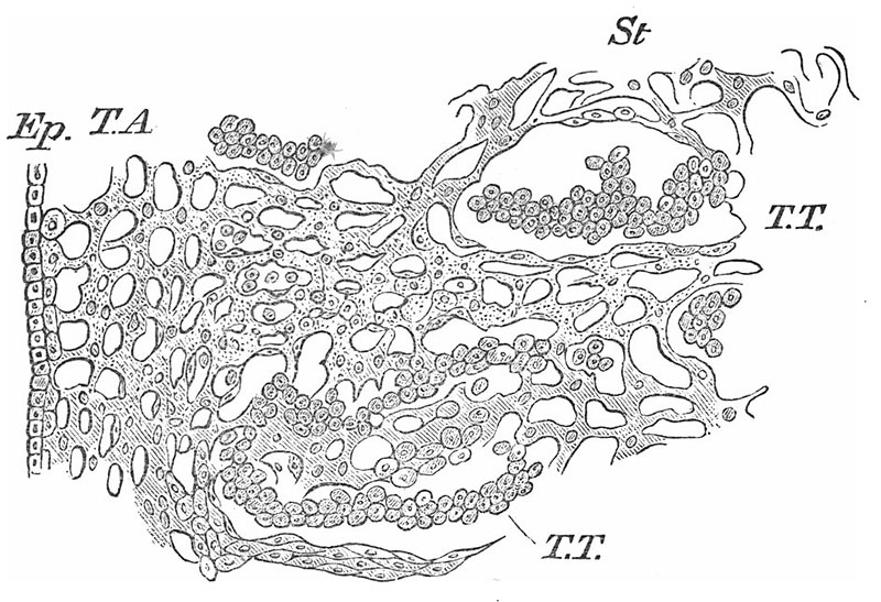
Fig. 41. Section through Testis at tenth week
To show developing seminal tubules and stroma.
Ep., epithelium ; T.A., tunica albuginea ; S., stroma; T.T., tubuli testis. x 250.== Lockwood1887b fig41.jpg
Reference
Lockwood CB. Development and transition of the testis, normal and abnormal. (1887) J Anat. 22(1): 38-77. PMID 17231729
Cite this page: Hill, M.A. (2024, April 18) Embryology Lockwood1887b fig41.jpg. Retrieved from https://embryology.med.unsw.edu.au/embryology/index.php/File:Lockwood1887b_fig41.jpg
- © Dr Mark Hill 2024, UNSW Embryology ISBN: 978 0 7334 2609 4 - UNSW CRICOS Provider Code No. 00098G
File history
Click on a date/time to view the file as it appeared at that time.
| Date/Time | Thumbnail | Dimensions | User | Comment | |
|---|---|---|---|---|---|
| current | 12:50, 14 April 2020 | | 800 × 547 (129 KB) | Z8600021 (talk | contribs) | contrast and size |
| 12:01, 14 April 2020 |  | 1,186 × 790 (184 KB) | Z8600021 (talk | contribs) | Lockwood1887b fig41.jpg |
You cannot overwrite this file.
File usage
The following page uses this file: