File:Liver-sinusoid colour cartoon.jpg: Difference between revisions
mNo edit summary |
mNo edit summary |
||
| (One intermediate revision by the same user not shown) | |||
| Line 5: | Line 5: | ||
:'''Links:''' [[Gastrointestinal Tract - Liver Development|Liver Development]] | [[Gastrointestinal Tract - Liver Histology|Liver Histology]] | [[Gastrointestinal Tract Development]] | |||
===Reference=== | ===Reference=== | ||
| Line 20: | Line 20: | ||
Modified from Figure 1. Journal.pbio.0030192.g001.png Modification - letter labels removed and full labels were added to the original diagram, colour added to B&W image. | Modified from Figure 1. Journal.pbio.0030192.g001.png Modification - letter labels removed and full labels were added to the original diagram, colour added to B&W image. | ||
{{Footer}} | |||
[[Category:Liver]] [[Category:Gastrointestinal Tract]] [[Category:Cartoon]] | [[Category:Liver]] [[Category:Gastrointestinal Tract]] [[Category:Cartoon]] | ||
Latest revision as of 07:45, 4 November 2015
Architecture of the Liver Sinusoid
- Liver sinusoids are lined by fenestrated endothelia and interspersed Kupffer cells, the resident macrophages of the liver.
- Stellate cells, the major producers of liver extracellular matrix, are located inside the narrow space of Disse, which is formed by the sinusoidal cell layer and cords of hepatocytes.
Reference
<pubmed>15901208</pubmed>| PLoS Biol.
Copyright
© 2005 Frevert et al. This is an open-access article distributed under the terms of the Creative Commons Attribution License, which permits unrestricted use, distribution, and reproduction in any medium, provided the original work is properly cited.
Citation: Frevert U, Engelmann S, Zougbédé S, Stange J, Ng B, et al. (2005) Intravital Observation of Plasmodium berghei Sporozoite Infection of the Liver. PLoS Biol 3(6): e192. doi:10.1371/journal.pbio.0030192
Modified from Figure 1. Journal.pbio.0030192.g001.png Modification - letter labels removed and full labels were added to the original diagram, colour added to B&W image.
Cite this page: Hill, M.A. (2024, April 19) Embryology Liver-sinusoid colour cartoon.jpg. Retrieved from https://embryology.med.unsw.edu.au/embryology/index.php/File:Liver-sinusoid_colour_cartoon.jpg
- © Dr Mark Hill 2024, UNSW Embryology ISBN: 978 0 7334 2609 4 - UNSW CRICOS Provider Code No. 00098G
File history
Click on a date/time to view the file as it appeared at that time.
| Date/Time | Thumbnail | Dimensions | User | Comment | |
|---|---|---|---|---|---|
| current | 09:19, 1 May 2010 | 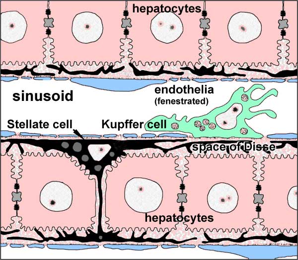 | 600 × 523 (64 KB) | S8600021 (talk | contribs) | Architecture of the Liver Sinusoid Liver sinusoids (S) are lined by fenestrated endothelia (EC) and interspersed Kupffer cells (KC), the resident macrophages of the liver. Stellate cells (SC), the major producers of liver ECM, are located inside the narr |
You cannot overwrite this file.
File usage
The following 3 pages use this file: