File:LewisFT1920 fig10-13.jpg
Original file (2,348 × 3,299 pixels, file size: 442 KB, MIME type: image/jpeg)
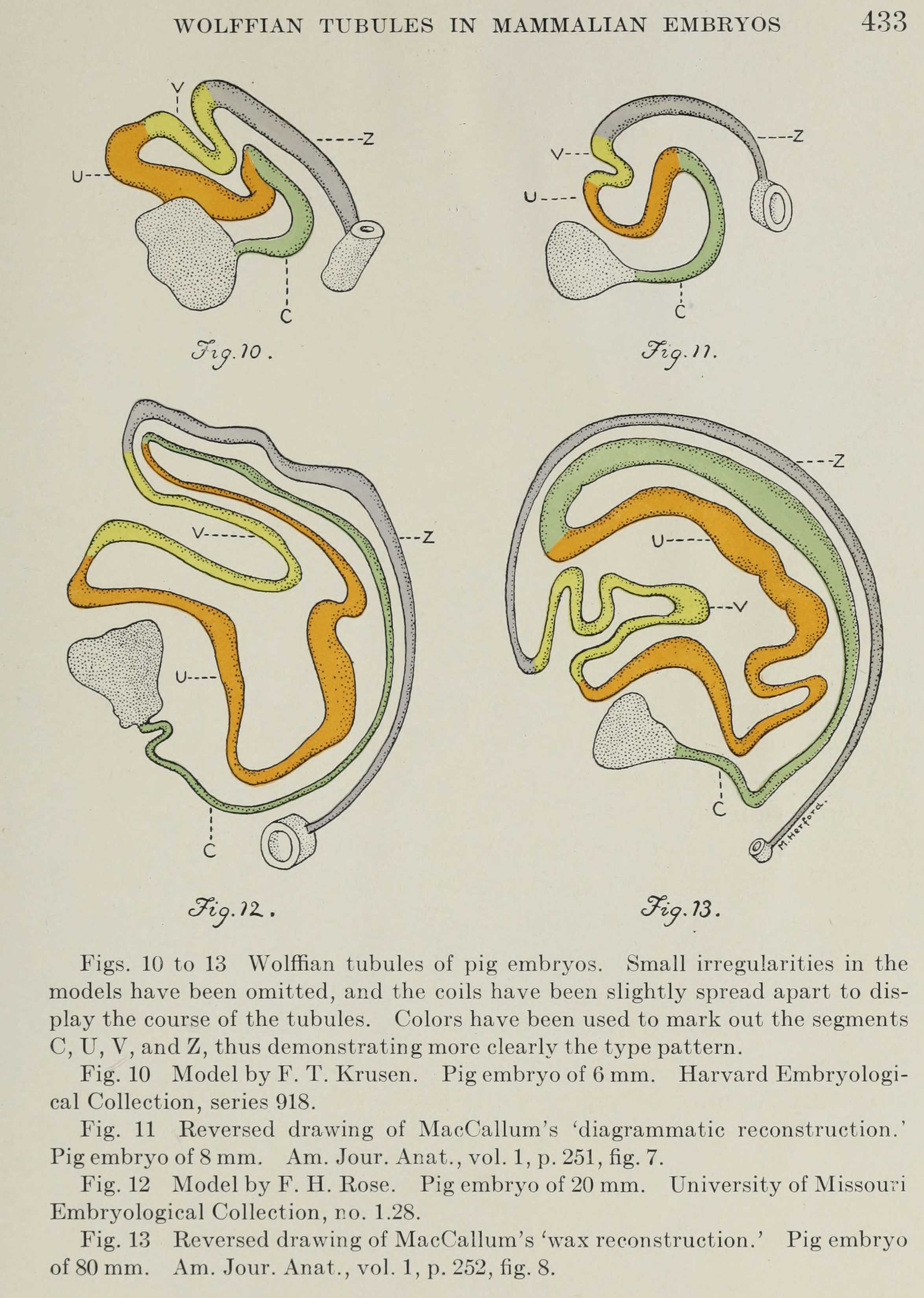
Fig. 10 Model by F. T. Krusen. Pig embryo of 6 mm. Harvard Embryological Collection, series 918.
Fig. 11 Reversed drawing of MacCallum's - diagrammatic reconstruction. Pig embryo of 8 mm. Am. Jour. Anat., vol. 1, p. 251, fig. 7.
Fig. 12 Model by F. H. Rose. Pig embryo of 20 mm. University of Missouri Embryological Collection, no. 1.28.
Fig. 13 Reversed drawing of MacCallum's - wax reconstruction. Pig embryo of 80mm. Am. Jour. Anat., vol. 1, p. 252, fig. 8.
<gallery> File:LewisFT1920 fig10.jpg|10 Pig embryo 6 mm File:LewisFT1920 fig11.jpg|11 Pig embryo 8 mm File:LewisFT1920 fig12.jpg|12 Pig embryo 20 mm File:LewisFT1920 fig13.jpg|13 Pig embryo 80 mm <gallery>
Reference
Lewis FT. The course of the Wolffian tubules in mammalian embryos. (1920) Amer. J Anat. 26(3): 423-436.
Cite this page: Hill, M.A. (2024, April 25) Embryology LewisFT1920 fig10-13.jpg. Retrieved from https://embryology.med.unsw.edu.au/embryology/index.php/File:LewisFT1920_fig10-13.jpg
- © Dr Mark Hill 2024, UNSW Embryology ISBN: 978 0 7334 2609 4 - UNSW CRICOS Provider Code No. 00098G
File history
Click on a date/time to view the file as it appeared at that time.
| Date/Time | Thumbnail | Dimensions | User | Comment | |
|---|---|---|---|---|---|
| current | 14:32, 9 August 2018 | | 2,348 × 3,299 (442 KB) | Z8600021 (talk | contribs) | ===Reference=== {{Ref-LewisFT1920}} {{Footer}} |
You cannot overwrite this file.
File usage
The following page uses this file: