File:Lewis1912-fig01.jpg
From Embryology
Size of this preview: 800 × 534 pixels. Other resolution: 1,000 × 668 pixels.
Original file (1,000 × 668 pixels, file size: 169 KB, MIME type: image/jpeg)
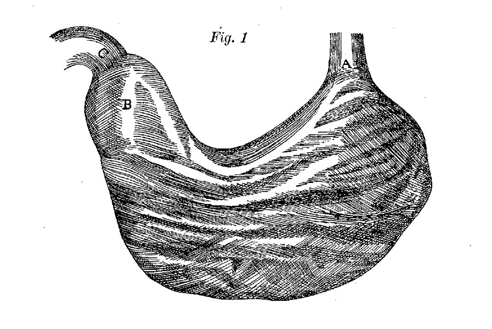
Fig, 1 Willis’s figure of the inverted stomach redrawn and reduced one-half
"A, Orificium sinistrum, sive os ventrieuli. B, Pylori Antrum, in que, Tunicae crassiores exist-unt. C, Orificium ejus, cuo Duodenum annectitur.”
| Historic Disclaimer - information about historic embryology pages |
|---|
 |
Reference
Lewis FT. The form of the stomach in human embryos with notes upon the nomenclature of the stomach. (1912) Amer. J Anat. 13(4): 477-503.
Cite this page: Hill, M.A. (2024, April 16) Embryology Lewis1912-fig01.jpg. Retrieved from https://embryology.med.unsw.edu.au/embryology/index.php/File:Lewis1912-fig01.jpg
- © Dr Mark Hill 2024, UNSW Embryology ISBN: 978 0 7334 2609 4 - UNSW CRICOS Provider Code No. 00098G
File history
Click on a date/time to view the file as it appeared at that time.
| Date/Time | Thumbnail | Dimensions | User | Comment | |
|---|---|---|---|---|---|
| current | 10:39, 25 April 2017 | | 1,000 × 668 (169 KB) | Z8600021 (talk | contribs) | {{Historic Disclaimer}} ===Reference=== {{Ref-Lewis1912}} {{Footer}} Category:StomachCategory:Historic EmbryologyCategory:1910's [[ |
You cannot overwrite this file.
File usage
The following page uses this file: