File:LattaTollman1937 fig12.jpg: Difference between revisions
From Embryology
({{LattaTollman1937 figures}}) |
mNo edit summary |
||
| Line 1: | Line 1: | ||
==Fig. 12. Mesoderm of the basal chorion== | |||
Most of the cells are fusiform with elongate nuclei containing scattered, finely divided chromatin. Occasional free spherical cells are to be found. Chorionic epithelium to the right with shrinkage space between it and the rnesoderm. X 600. | |||
{{LattaTollman1937 figures}} | {{LattaTollman1937 figures}} | ||
Latest revision as of 12:58, 26 February 2017
Fig. 12. Mesoderm of the basal chorion
Most of the cells are fusiform with elongate nuclei containing scattered, finely divided chromatin. Occasional free spherical cells are to be found. Chorionic epithelium to the right with shrinkage space between it and the rnesoderm. X 600.
- Links: fig 1 | fig 2 | fig 3 | fig 4 | fig 5 | fig 6 | fig 7 | fig 8 | fig 9 | fig 10 | fig 11 | fig 12 | fig 13 | fig 14 | fig 15 | plate 1 | plate 2 | plate 3 | 1937 Latta Tollman | Historic Papers
Reference
Latta JS. and Tollman JP. An early stage of human implantation. (1937) Anat. Rec. 69(4): 443-463.
Cite this page: Hill, M.A. (2024, April 19) Embryology LattaTollman1937 fig12.jpg. Retrieved from https://embryology.med.unsw.edu.au/embryology/index.php/File:LattaTollman1937_fig12.jpg
- © Dr Mark Hill 2024, UNSW Embryology ISBN: 978 0 7334 2609 4 - UNSW CRICOS Provider Code No. 00098G
File history
Click on a date/time to view the file as it appeared at that time.
| Date/Time | Thumbnail | Dimensions | User | Comment | |
|---|---|---|---|---|---|
| current | 12:56, 26 February 2017 | 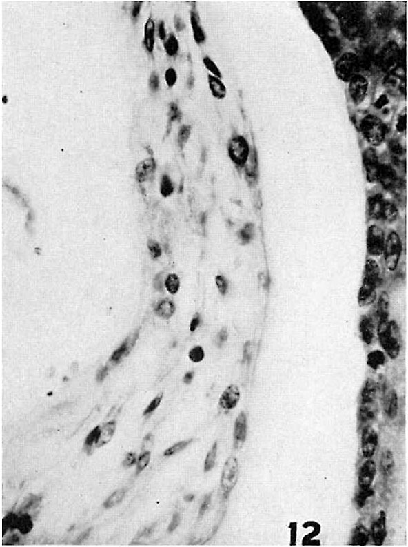 | 592 × 794 (85 KB) | Z8600021 (talk | contribs) | {{LattaTollman1937 figures}} |
You cannot overwrite this file.
File usage
The following page uses this file: