File:LattaTollman1937 fig01.jpg
Original file (1,000 × 1,281 pixels, file size: 416 KB, MIME type: image/jpeg)
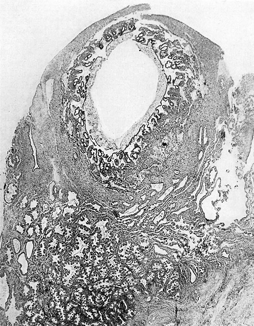
Fig. 1. A section near the center of the implanted ovum
The enlargement of the uterine glands and the numerous papillary folds are clearly shown. Note the comparative absence of glands in the more compact tissue immediately surrounding the implantation cavity and also compactness of the glandular tissue in the region immediately underneath the ovum. X 18.
- Links: fig 1 | fig 2 | fig 3 | fig 4 | fig 5 | fig 6 | fig 7 | fig 8 | fig 9 | fig 10 | fig 11 | fig 12 | fig 13 | fig 14 | fig 15 | plate 1 | plate 2 | plate 3 | 1937 Latta Tollman | Historic Papers
Reference
Latta JS. and Tollman JP. An early stage of human implantation. (1937) Anat. Rec. 69(4): 443-463.
Cite this page: Hill, M.A. (2024, April 25) Embryology LattaTollman1937 fig01.jpg. Retrieved from https://embryology.med.unsw.edu.au/embryology/index.php/File:LattaTollman1937_fig01.jpg
- © Dr Mark Hill 2024, UNSW Embryology ISBN: 978 0 7334 2609 4 - UNSW CRICOS Provider Code No. 00098G
File history
Click on a date/time to view the file as it appeared at that time.
| Date/Time | Thumbnail | Dimensions | User | Comment | |
|---|---|---|---|---|---|
| current | 12:01, 26 February 2017 | | 1,000 × 1,281 (416 KB) | Z8600021 (talk | contribs) | |
| 11:59, 26 February 2017 |  | 1,327 × 1,901 (568 KB) | Z8600021 (talk | contribs) |
You cannot overwrite this file.
File usage
The following page uses this file: