File:Kuntz1920 plate07.jpg
Original file (1,280 × 1,836 pixels, file size: 622 KB, MIME type: image/jpeg)
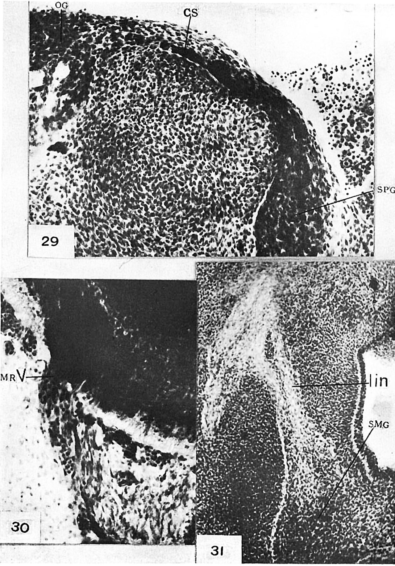
Plate 7
29 Human embryo, 14 mm. in length, 144*5—2—2 x 240. Sagittal section showing cellular ramus (CS) connecting otic (O0) and sphenopalatine (SPG) ganglia.
30 Human embryo, 14.5 mm. in length, 1267—8—2—3 x 3l5. Section through motor root of trigcminal nerve (MRV) showing migrant medullary cells.
31 Human embryo, 13 mm. in length, 485—13—8—3 x 120. Section showing primordium of submaxillary ganglion (SMG). lain, lingual nerve.
Reference
Kuntz A. The development of the sympathetic nervous system in man. (1920) J. Comp. Neurol. 32(2): 173-229.
Cite this page: Hill, M.A. (2024, April 16) Embryology Kuntz1920 plate07.jpg. Retrieved from https://embryology.med.unsw.edu.au/embryology/index.php/File:Kuntz1920_plate07.jpg
- © Dr Mark Hill 2024, UNSW Embryology ISBN: 978 0 7334 2609 4 - UNSW CRICOS Provider Code No. 00098G
File history
Click on a date/time to view the file as it appeared at that time.
| Date/Time | Thumbnail | Dimensions | User | Comment | |
|---|---|---|---|---|---|
| current | 17:40, 28 March 2017 | | 1,280 × 1,836 (622 KB) | Z8600021 (talk | contribs) | |
| 17:40, 28 March 2017 | 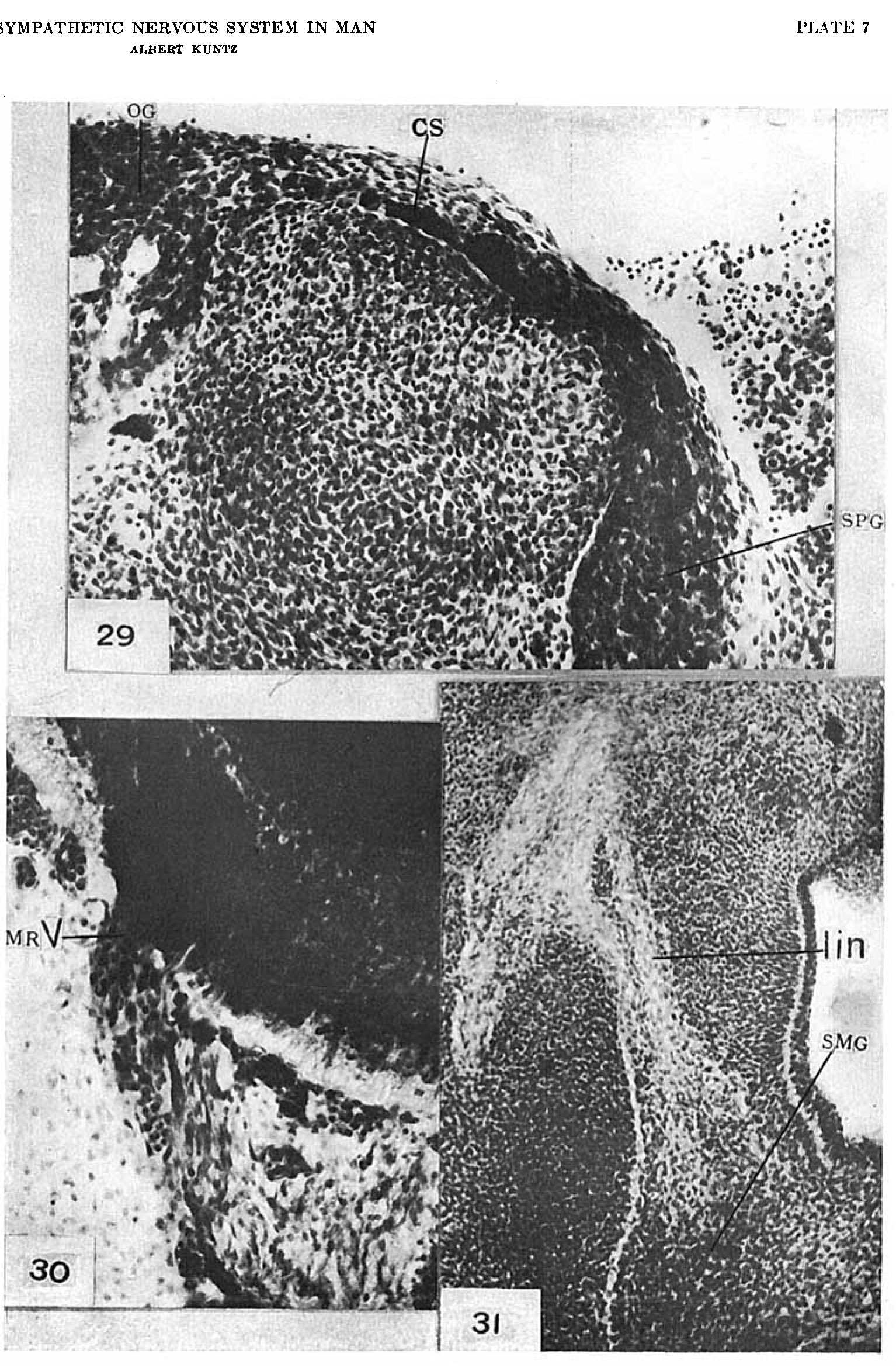 | 1,534 × 2,345 (791 KB) | Z8600021 (talk | contribs) | ===Plate 7=== 29 Human embryo, 14 mm. in length, 144*5—2—2 x 240. Sagittal section showing cellular ramus (CS) connecting otic (O0) and sphenopalatine (SPG) ganglia. 30 Human embryo, 14.5 mm. in length, [[:Category:Carnegie Embryo 1267|'''1267'''... |
You cannot overwrite this file.
File usage
The following page uses this file: