File:Kollmann761.jpg
Kollmann761.jpg (641 × 455 pixels, file size: 45 KB, MIME type: image/jpeg)
- This text is a Google translate computer generated translation and may contain many errors.
Images from - Atlas of the Development of Man (Volume 2)
(Handatlas der entwicklungsgeschichte des menschen)
- Kollmann Atlas 2: Gastrointestinal | Respiratory | Urogenital | Cardiovascular | Neural | Integumentary | Smell | Vision | Hearing | Kollmann Atlas 1 | Kollmann Atlas 2 | Julius Kollmann
- Links: Julius Kollman | Atlas Vol.1 | Atlas Vol.2 | Embryology History
| Historic Disclaimer - information about historic embryology pages |
|---|
 |
Reference
Kollmann JKE. Atlas of the Development of Man (Handatlas der entwicklungsgeschichte des menschen). (1907) Vol.1 and Vol. 2. Jena, Gustav Fischer. (1898).
Cite this page: Hill, M.A. (2024, April 18) Embryology Kollmann761.jpg. Retrieved from https://embryology.med.unsw.edu.au/embryology/index.php/File:Kollmann761.jpg
- © Dr Mark Hill 2024, UNSW Embryology ISBN: 978 0 7334 2609 4 - UNSW CRICOS Provider Code No. 00098G
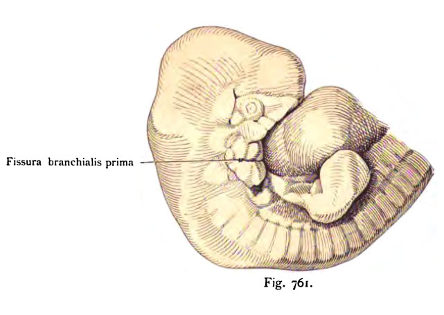
Fig. 761. Oberkörper eines menschlichen Embryo von 10,4 mm Scheitelsteifi-
länse.
(Alter 4 Wochen.) Der Kopf ist senkrecht orientiert damit seine Besichtigung um
so leichter sei.
(Nach G. Retzius.)
Die I. Kiemenspalte ist bezeichnet. Oral von ihr befindet sich der Mandi- bularbogen mit Aurikularhöckern, CoUiculi auriculares, aboral der IL Kiemen- bogen oder Hyoidbogen, gleichfalls mit Aurikularhöckern versehen. Dorsal schimmert die Vesiculä auditiva hindurch. Dieser Embryo ist ausgezeichnet erhalten, die Aurikularhöcker besitzen scharfe Formen wie an einem Basler Embryo des nämlichen Alters. Die folgenden Figuren geben die weiteren Ver- änderungen dieser Aurikularhöcker.
File history
Click on a date/time to view the file as it appeared at that time.
| Date/Time | Thumbnail | Dimensions | User | Comment | |
|---|---|---|---|---|---|
| current | 12:41, 21 October 2011 | | 641 × 455 (45 KB) | S8600021 (talk | contribs) | {{Kollmann1907}} Category:Hearing Fig. 761. Oberkörper eines menschlichen Embryo von 10,4 mm Scheitelsteifi- länse. (Alter 4 Wochen.) Der Kopf ist senkrecht orientiert damit seine Besichtigung um so leichter sei. (Nach G. Retzius.) Die |
You cannot overwrite this file.
File usage
The following page uses this file: