File:Kollmann757.jpg
Original file (807 × 697 pixels, file size: 98 KB, MIME type: image/jpeg)
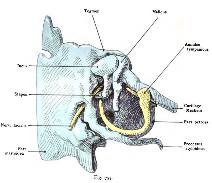
Fig. 757. Middle ear of a human fetus 8 cm CRL
The ear drum is removed.
(Anatomical Collection in Basel.)
In the cavity are the three ossicles auditus. The hammer is in connection with the Meckel's cartilage. Behind the ossicles auditus is the Paries labyrinthica. Behind the root of the styloid process stylomastoid foramen is found with the N. facial nerve, whose course in the cavum tympani is still a far stretch freely. The tympanic annulus (yellow because of covering bone) limits the Paries membranous, consisting in the absence of the eardrum, the figure only from the open top cartilage ring.
Comparisons for the evolution of the tympanic cavity is found in the osteology section: Cranium the Fig. 262 and Fig. 264.
- This text is a Google translate computer generated translation and may contain many errors.
Images from - Atlas of the Development of Man (Volume 2)
(Handatlas der entwicklungsgeschichte des menschen)
- Kollmann Atlas 2: Gastrointestinal | Respiratory | Urogenital | Cardiovascular | Neural | Integumentary | Smell | Vision | Hearing | Kollmann Atlas 1 | Kollmann Atlas 2 | Julius Kollmann
- Links: Julius Kollman | Atlas Vol.1 | Atlas Vol.2 | Embryology History
| Historic Disclaimer - information about historic embryology pages |
|---|
 |
Reference
Kollmann JKE. Atlas of the Development of Man (Handatlas der entwicklungsgeschichte des menschen). (1907) Vol.1 and Vol. 2. Jena, Gustav Fischer. (1898).
Cite this page: Hill, M.A. (2024, April 24) Embryology Kollmann757.jpg. Retrieved from https://embryology.med.unsw.edu.au/embryology/index.php/File:Kollmann757.jpg
- © Dr Mark Hill 2024, UNSW Embryology ISBN: 978 0 7334 2609 4 - UNSW CRICOS Provider Code No. 00098G
Fig. 757. Mitteiohrraum^ Cavum tympani emes menschlichen Fetus
von 8 cm Scheitelsteißlänge. Das Trommelfell ist entfernt.
(Anatomische Sammlung in Basel.)
Im Cavum befinden sich die drei Ossicula auditus. Der Hammer steht in Verbindung mit dem M eck eischen Knorpel. Hinter den Ossicula auditus liegt die Paries labyrinthica. Hinter der Wurzel des Processus styloideus be- findet sich das Foramen stylomastoideum mit dem N. facialis, dessen Verlauf im Cavum tympani noch eine Strecke weit frei liegt. Der Annulus tympanicus (gelb weil ein Belegknochen) begrenzt die Paries membranacea, die beim Fehlen des Trommelfelles in der Abbildung nur aus dem nach oben offenen Knorpel- ring besteht.
Vergleiche für die Entwicklungsgeschichte des Cavum tympani auch in dem Abschnitt Osteologie: Cranium die Figg. 262 und 264.
File history
Click on a date/time to view the file as it appeared at that time.
| Date/Time | Thumbnail | Dimensions | User | Comment | |
|---|---|---|---|---|---|
| current | 12:39, 21 October 2011 | | 807 × 697 (98 KB) | S8600021 (talk | contribs) | {{Kollmann1907}} Category:Hearing Fig. 757. Mitteiohrraum^ Cavum tympani emes menschlichen Fetus von 8 cm Scheitelsteißlänge. Das Trommelfell ist entfernt. (Anatomische Sammlung in Basel.) Im Cavum befinden sich die drei Ossicula auditus. |
You cannot overwrite this file.
File usage
The following page uses this file: