File:Kollmann756.jpg
Original file (820 × 489 pixels, file size: 67 KB, MIME type: image/jpeg)
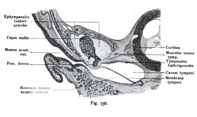
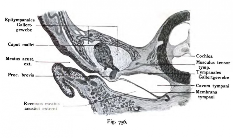
Fig. 756. Coronal section through the tympanic region of human fetus 11 cm CRL
(End of 4th month) (After Harnmar.)
The section passes through the outer ear, the ear canal, the recessus meatus and the tympanic cavity and meets the ear capsule in the region of the cochlea. The cavum tympani is a form which is shown in Fig 755 as full screen. The epitympanale area does not yet exist, but is later seen by the position taken by the average hammer head, the bottom of the cartilaginous ear canal shows a section, it corresponds to the primary auditory canal and a membranous portion, which goes into the ear gangrecessus. The recessus meatus acustici externi is highlighted, approached from the ear canal plate (lamina epithelialis meatus), the above is shown in Fig 753 and Fig 754.
- This text is a Google translate computer generated translation and may contain many errors.
Images from - Atlas of the Development of Man (Volume 2)
(Handatlas der entwicklungsgeschichte des menschen)
- Kollmann Atlas 2: Gastrointestinal | Respiratory | Urogenital | Cardiovascular | Neural | Integumentary | Smell | Vision | Hearing | Kollmann Atlas 1 | Kollmann Atlas 2 | Julius Kollmann
- Links: Julius Kollman | Atlas Vol.1 | Atlas Vol.2 | Embryology History
| Historic Disclaimer - information about historic embryology pages |
|---|
 |
Reference
Kollmann JKE. Atlas of the Development of Man (Handatlas der entwicklungsgeschichte des menschen). (1907) Vol.1 and Vol. 2. Jena, Gustav Fischer. (1898).
Cite this page: Hill, M.A. (2024, April 18) Embryology Kollmann756.jpg. Retrieved from https://embryology.med.unsw.edu.au/embryology/index.php/File:Kollmann756.jpg
- © Dr Mark Hill 2024, UNSW Embryology ISBN: 978 0 7334 2609 4 - UNSW CRICOS Provider Code No. 00098G
Fig. 756. Frontalschnitt durch die PaykenhShle eines Menschenfetus von
11 cm Scheitelsteifilänse.
(Ende des 4. Monats.) (Nach Harn mar.)
Der Schnitt geht durch das äußere Ohr, den äußeren Gehörgang, den Recessus meatus, und trifft das Cavum tympani und die Ohrkapsel im Bereich der Cochlea. Das Cavum tympani zeigt eine Form, welche in der Fig. 755 als Vollbild dargestellt ist. Der epitympanale Raum besteht noch nicht, doch ist die spätere Lage ersichtlich durch den vom Schnitt getroffenen Hammerkopf; der Boden des Gehörganges zeigt einen knorpeligen Abschnitt, er entspricht dem primären Gehörgang und einem häutigen Abschnitt, der in den Gehör- gangrecessus übergeht. Der Recessus meatus acustici externi ist hervorge- gangen aus der Gehörgangsplatte (Lamina epithelialis meatus), die oben in den Figg. 753 und 754 dargestellt ist.
File history
Click on a date/time to view the file as it appeared at that time.
| Date/Time | Thumbnail | Dimensions | User | Comment | |
|---|---|---|---|---|---|
| current | 12:39, 21 October 2011 | | 820 × 489 (67 KB) | S8600021 (talk | contribs) | {{Kollmann1907}} Category:Hearing Fig. 756. Frontalschnitt durch die PaykenhShle eines Menschenfetus von 11 cm Scheitelsteifilänse. (Ende des 4. Monats.) (Nach Harn mar.) Der Schnitt geht durch das äußere Ohr, den äußeren Gehörgang, de |
You cannot overwrite this file.
File usage
The following page uses this file: