File:Kollmann755.jpg

Original file (814 × 457 pixels, file size: 59 KB, MIME type: image/jpeg)
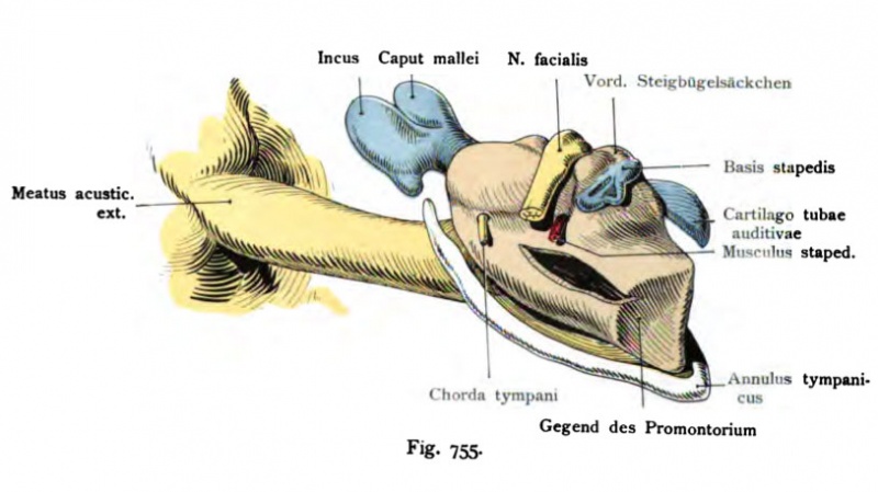
Fig. 755. Acoustic region in a human fetus of 22.5 cm 5th Month
External acoustic meatus (yellow), tympanic cavity (red), ossicles auditus (blue)
(Reconstruction) View from behind. The figure was slightly simplified.
(After Harn mar.)
- This text is a Google translate computer generated translation and may contain many errors.
Images from - Atlas of the Development of Man (Volume 2)
(Handatlas der entwicklungsgeschichte des menschen)
- Kollmann Atlas 2: Gastrointestinal | Respiratory | Urogenital | Cardiovascular | Neural | Integumentary | Smell | Vision | Hearing | Kollmann Atlas 1 | Kollmann Atlas 2 | Julius Kollmann
- Links: Julius Kollman | Atlas Vol.1 | Atlas Vol.2 | Embryology History
| Historic Disclaimer - information about historic embryology pages |
|---|
 |
Reference
Kollmann JKE. Atlas of the Development of Man (Handatlas der entwicklungsgeschichte des menschen). (1907) Vol.1 and Vol. 2. Jena, Gustav Fischer. (1898).
Cite this page: Hill, M.A. (2024, April 23) Embryology Kollmann755.jpg. Retrieved from https://embryology.med.unsw.edu.au/embryology/index.php/File:Kollmann755.jpg
- © Dr Mark Hill 2024, UNSW Embryology ISBN: 978 0 7334 2609 4 - UNSW CRICOS Provider Code No. 00098G
Fig. 755. Meatus acusticus externus (gelb), Cavum tympani (rot), Ossicula
auditus (blau)
bei einem menschlichen Fetus von 22,5 cm. 5. Monat. (Rekonstruktion.) Ansicht
von hinten. Die Figur wurde etwas vereinfacht.
(Nach Harn mar.)
Die knorpelige Ohrkapsel ist entfernt, so daß in erster. Linie das rot an- gelegte Cavum tympani auffällt. Zur Orientierung ist es wertvoll, die Lage der Gehörknöchelchen zu beachten, und den Annulus tympanicus, der den Rand des Trommelfells umspannt. Das Cavum tympani sieht aus wie ein unregel- mäßiger Polster, der sich vom Trommelfell erhebt. Es existiert nur die An- lage des eigentlichen Cavum ; Aditus ad antrum und Antrum fehlen noch. Um den Eindruck des Cavum hervorzurufen, ist ein spaltförmiges Stück ausge- schnitten worden. Man sieht in das Cavum hinein, dessen Epithelgjenze durch die Rekonstruktion festgestellt wurde.
File history
Click on a date/time to view the file as it appeared at that time.
| Date/Time | Thumbnail | Dimensions | User | Comment | |
|---|---|---|---|---|---|
| current | 12:38, 21 October 2011 | | 814 × 457 (59 KB) | S8600021 (talk | contribs) | {{Kollmann1907}} Category:Hearing Fig. 755. Meatus acusticus externus (gelb), Cavum tympani (rot), Ossicula auditus (blau) bei einem menschlichen Fetus von 22,5 cm. 5. Monat. (Rekonstruktion.) Ansicht von hinten. Die Figur wurde etwas vereinfa |
You cannot overwrite this file.
File usage
The following page uses this file: