File:Kollmann754.jpg

Original file (802 × 577 pixels, file size: 71 KB, MIME type: image/jpeg)
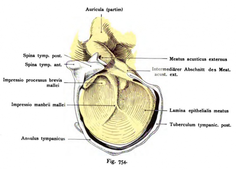
Fig. 754. Auditory meatus externus from a human fetus 22.5 cm (5th month)
Reconstruction view from above and inside.
(After Harn mar.)
The external acoustic meatus consists of three sections: i. an outer, which corresponds to the primary means of the external auditory canal Kiemehtasche; in it later occurred to hair, glands, and cartilage; second an inner broad, rounded section, which is limited by the tympanum (the ear canal plate, lamina epithelialis meatus), 3rd a small intermediate section located laterally of the plate, and the hair remains drtisenfrei. The ear canal plate is now develops into a roundish, thin, solid disc, which is connected at its upper edge to the far narrower primary ear canal as with a stick. In the 7th Month split this record. The resulting column occurs with the clearing of the outer ear canal in connection with which the definitive ear canal is formed.
- This text is a Google translate computer generated translation and may contain many errors.
Images from - Atlas of the Development of Man (Volume 2)
(Handatlas der entwicklungsgeschichte des menschen)
- Kollmann Atlas 2: Gastrointestinal | Respiratory | Urogenital | Cardiovascular | Neural | Integumentary | Smell | Vision | Hearing | Kollmann Atlas 1 | Kollmann Atlas 2 | Julius Kollmann
- Links: Julius Kollman | Atlas Vol.1 | Atlas Vol.2 | Embryology History
| Historic Disclaimer - information about historic embryology pages |
|---|
| Pages where the terms "Historic" (textbooks, papers, people, recommendations) appear on this site, and sections within pages where this disclaimer appears, indicate that the content and scientific understanding are specific to the time of publication. This means that while some scientific descriptions are still accurate, the terminology and interpretation of the developmental mechanisms reflect the understanding at the time of original publication and those of the preceding periods, these terms, interpretations and recommendations may not reflect our current scientific understanding. (More? Embryology History | Historic Embryology Papers) |
Reference
Kollmann JKE. Atlas of the Development of Man (Handatlas der entwicklungsgeschichte des menschen). (1907) Vol.1 and Vol. 2. Jena, Gustav Fischer. (1898).
Cite this page: Hill, M.A. (2024, April 23) Embryology Kollmann754.jpg. Retrieved from https://embryology.med.unsw.edu.au/embryology/index.php/File:Kollmann754.jpg
- © Dr Mark Hill 2024, UNSW Embryology ISBN: 978 0 7334 2609 4 - UNSW CRICOS Provider Code No. 00098G
Fig. 754. Äußerer Qehörgans, Meatus acusticus extemus von einem menschlichen Fetus von 22,5 cm.
(5. Monat.) Rekonstruktion.. Ansicht von oben und innen.
(Nach einem Modell von Harn mar.)
Der Meatus acusticus externus besteht aus drei Abschnitten: i. einem äußeren, welcher dem primären Gehörgang d. h. der äußeren Kiemehtasche entspricht; in ihm treten später Haare, Drüsen und Knorpel auf; 2. einem inneren breiten, rundlichen Abschnitt, der vom Paukenfell begrenzt wird (die Gehörgangplatte, Lamina epithelialis meatus); 3. einem kleinen intermediären Abschnitt, lateral von der Platte gelegen, der haar- und drtisenfrei bleibt. Die Gehörgangplatte ist jetzt zu einer rundlichen, dünnen, soliden Scheibe entwickelt, welche an ihrem oberen Rande mit dem weit schmäleren primären Gehörgang wie mit einem Stiel zusammenhängt. Im 7. Monat spaltet sich diese Platte. Die entstandene Spalte tritt mit der Lichtung des äußeren Gehörgangs in Verbindung, womit der definitive Gehörgang gebildet ist.
File history
Click on a date/time to view the file as it appeared at that time.
| Date/Time | Thumbnail | Dimensions | User | Comment | |
|---|---|---|---|---|---|
| current | 12:38, 21 October 2011 |  | 802 × 577 (71 KB) | S8600021 (talk | contribs) | {{Kollmann1907}} Category:Hearing Fig. 754. Äußerer Qehörgans, Meatus acusticus extemus von einem mensch- lichen Fetus von 22,5 cm. (5. Monat.) Rekonstruktion.. Ansicht von oben und innen. (Nach einem Modell von Harn mar.) Der Meatus ac |
You cannot overwrite this file.
File usage
The following page uses this file:
