File:Kollmann753.jpg
Kollmann753.jpg (734 × 380 pixels, file size: 41 KB, MIME type: image/jpeg)
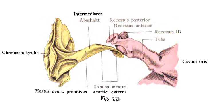
Fig. 753. Tubo-tympanic recess and external ear of a human fetus of 31 mm CRL
(Middle of the 3rd month.)
View from in front.
(After Harn mar.)
The primary tympanic cavity provides an environment with the primary tube tube is elongated plate, hence the name "tubo-tympanic." There extends from the sides of the dorsal pharyngeal roof gradually extending. The boundary between the primary and middle ear cavity of the primary tube is marked by an asterisk. One recognizes the primary tympanic cavity, moreover to the increase in the width and the appearance of the recesses. the primary ear forms a cylindrical tube with a funnel which short-shaped piece at the beginning of the ear pit begins. This is followed by the actual ear canal, which passes through an intermediate section in the ear canal plate, lamina epithelialis meatus. This plate is in the beginning in a solid form that is later cleared. These solid ear canal plate grows in the 4th and 5 Month to a disk.
- This text is a Google translate computer generated translation and may contain many errors.
Images from - Atlas of the Development of Man (Volume 2)
(Handatlas der entwicklungsgeschichte des menschen)
- Kollmann Atlas 2: Gastrointestinal | Respiratory | Urogenital | Cardiovascular | Neural | Integumentary | Smell | Vision | Hearing | Kollmann Atlas 1 | Kollmann Atlas 2 | Julius Kollmann
- Links: Julius Kollman | Atlas Vol.1 | Atlas Vol.2 | Embryology History
| Historic Disclaimer - information about historic embryology pages |
|---|
| Pages where the terms "Historic" (textbooks, papers, people, recommendations) appear on this site, and sections within pages where this disclaimer appears, indicate that the content and scientific understanding are specific to the time of publication. This means that while some scientific descriptions are still accurate, the terminology and interpretation of the developmental mechanisms reflect the understanding at the time of original publication and those of the preceding periods, these terms, interpretations and recommendations may not reflect our current scientific understanding. (More? Embryology History | Historic Embryology Papers) |
Reference
Kollmann JKE. Atlas of the Development of Man (Handatlas der entwicklungsgeschichte des menschen). (1907) Vol.1 and Vol. 2. Jena, Gustav Fischer. (1898).
Cite this page: Hill, M.A. (2024, April 25) Embryology Kollmann753.jpg. Retrieved from https://embryology.med.unsw.edu.au/embryology/index.php/File:Kollmann753.jpg
- © Dr Mark Hill 2024, UNSW Embryology ISBN: 978 0 7334 2609 4 - UNSW CRICOS Provider Code No. 00098G
Fig. 753. Reclites tubo-tympanales Rolir und recliter äußerer Qeliörgang
von einem menschlichen Fetus von 31 mm Scheitelsteißlänge. (Mitte des 3. Monats.)
Ansicht von vom.
(Nach Harn mar.)
Die primäre Paukenhöhle stellt zusammen mit der primären Tube ein längliches plattes Rohr dar, daher die Bezeichnung „tubo-tympanal". Es er- streckt sich von den Seitenteilen des Schlunddaches dorsalwärts allmählich sich erweiternd. Die Grenze zwischen der primären Paukenhöhle und der primären Tube ist durch * bezeichnet. Man erkennt die primäre Paukenhöhle überdies an der Zunahme in der Breite und an dem Auftreten der Rezesse. Der primäre Gehörgang bildet ein zylindrisches Rohr, welches mit einem kurzen trichter- förmigen Anfangsstück an der Ohrmuschelgrube beginnt. Darauf folgt der eigentliche Gehörgang, der durch einen intermediären Abschnitt in die Gehör- gangsplatte, Lamina epithelialis meatus übergeht. Diese Platte ist im Anfange an eme solide Bildung, ihre Lichtung entsteht erst später. Diese solide Gehör- gangsplatte wächst im 4. und 5. Monat zu einer Scheibe aus.
File history
Click on a date/time to view the file as it appeared at that time.
| Date/Time | Thumbnail | Dimensions | User | Comment | |
|---|---|---|---|---|---|
| current | 12:37, 21 October 2011 |  | 734 × 380 (41 KB) | S8600021 (talk | contribs) | {{Kollmann1907}} Category:Hearing Fig. 753. Reclites tubo-tympanales Rolir und recliter äußerer Qeliörgang von einem menschlichen Fetus von 31 mm Scheitelsteißlänge. (Mitte des 3. Monats.) Ansicht von vom. (Nach Harn mar.) Die primä |
You cannot overwrite this file.
File usage
The following page uses this file:

