File:Kollmann751.jpg
Kollmann751.jpg (691 × 348 pixels, file size: 34 KB, MIME type: image/jpeg)
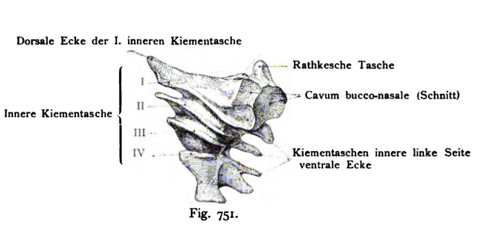
Fig. 751. Inner pharyngeal pouchs of a rabbit embryo of ?? days (5.3 mm in length)
60 times magnified reconstruction. (According to Piersol.)
The expansion of the four inner gill pockets from the ventral to dorsal surface of the embryo is visible in the form of narrow columns, which proceed from the foregut and terminate at the closing plate. This figure helps to understand the previous Fig. 750 and the following Fig. 752 figures. The results presented in two sections lie ventral to the front surface of the foregut.
(gill pockets = pharyngeal pouch)
- This text is a Google translate computer generated translation and may contain many errors.
Images from - Atlas of the Development of Man (Volume 2)
(Handatlas der entwicklungsgeschichte des menschen)
- Kollmann Atlas 2: Gastrointestinal | Respiratory | Urogenital | Cardiovascular | Neural | Integumentary | Smell | Vision | Hearing | Kollmann Atlas 1 | Kollmann Atlas 2 | Julius Kollmann
- Links: Julius Kollman | Atlas Vol.1 | Atlas Vol.2 | Embryology History
| Historic Disclaimer - information about historic embryology pages |
|---|
 |
Reference
Kollmann JKE. Atlas of the Development of Man (Handatlas der entwicklungsgeschichte des menschen). (1907) Vol.1 and Vol. 2. Jena, Gustav Fischer. (1898).
Cite this page: Hill, M.A. (2024, April 18) Embryology Kollmann751.jpg. Retrieved from https://embryology.med.unsw.edu.au/embryology/index.php/File:Kollmann751.jpg
- © Dr Mark Hill 2024, UNSW Embryology ISBN: 978 0 7334 2609 4 - UNSW CRICOS Provider Code No. 00098G
Fig. 751. Innere Kiementaschen eines Kaninchenembryo
von loVa Tagen (5,3 mm Länge) darstellend. 60 mal vergr. Rekonstruktion.
(Nach Piersol.)
Die Ausdehnung der vier inneren Kiementaschen von der ventralen bis zur dorsalen Fläche des Embryo ist erkennbar in Form schmaler Spalten, welche von dem Kopfdarm ausgehen und an der Verschlußplatte endigen. Diese Figur hilft die vorhergehende Fig. 750 und die nachfolgende (Fig. 752) richtig auffassen. Die in beiden dargestellten Schnitte liegen ventral zur vorder- fläche des Kopfdarms.
File history
Click on a date/time to view the file as it appeared at that time.
| Date/Time | Thumbnail | Dimensions | User | Comment | |
|---|---|---|---|---|---|
| current | 12:35, 21 October 2011 | | 691 × 348 (34 KB) | S8600021 (talk | contribs) | {{Kollmann1907}} Category:Hearing Fig. 751. Innere Kiementaschen eines Kaninchenembryo von loVa Tagen (5,3 mm Länge) darstellend. 60 mal vergr. Rekonstruktion. (Nach Piersol.) Die Ausdehnung der vier inneren Kiementaschen von der ventralen b |
You cannot overwrite this file.
File usage
The following page uses this file: