File:Kollmann750.jpg
Kollmann750.jpg (640 × 433 pixels, file size: 37 KB, MIME type: image/jpeg)
- This text is a Google translate computer generated translation and may contain many errors.
Images from - Atlas of the Development of Man (Volume 2)
(Handatlas der entwicklungsgeschichte des menschen)
- Kollmann Atlas 2: Gastrointestinal | Respiratory | Urogenital | Cardiovascular | Neural | Integumentary | Smell | Vision | Hearing | Kollmann Atlas 1 | Kollmann Atlas 2 | Julius Kollmann
- Links: Julius Kollman | Atlas Vol.1 | Atlas Vol.2 | Embryology History
| Historic Disclaimer - information about historic embryology pages |
|---|
 |
Reference
Kollmann JKE. Atlas of the Development of Man (Handatlas der entwicklungsgeschichte des menschen). (1907) Vol.1 and Vol. 2. Jena, Gustav Fischer. (1898).
Cite this page: Hill, M.A. (2024, April 24) Embryology Kollmann750.jpg. Retrieved from https://embryology.med.unsw.edu.au/embryology/index.php/File:Kollmann750.jpg
- © Dr Mark Hill 2024, UNSW Embryology ISBN: 978 0 7334 2609 4 - UNSW CRICOS Provider Code No. 00098G
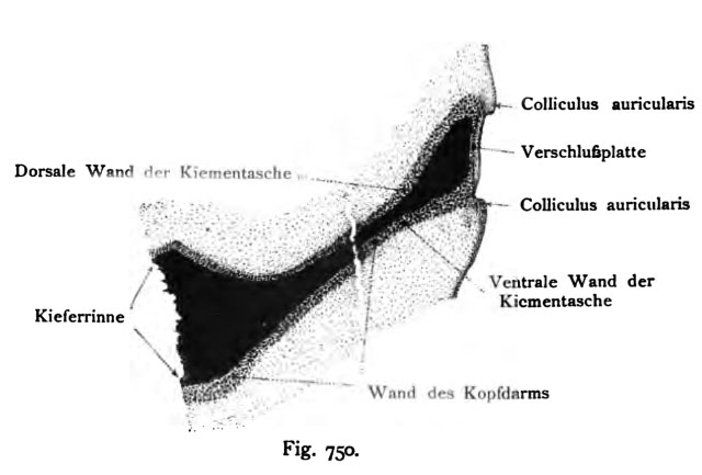
Fig. 750. Erste innere Kiementasclie eines menschlichen Embryo
von 10,2 mm Nackensteißlänge. 125 mal vergr. Dort wo der Riß in der Abbil- dung sich befindet, wurde ein Stück der Taschenlänge ausgeschaltet.
(Anatomische Sammlung in Basel)
Die Figur zeigt die große Ausdehnung der inneren Tasche, aus der je- doch nur die ventrale Abteilung der Trommelhöhle hervorgeht. Die Tuba auditiva fehlt noch, sie bildet sich später im Anschluß an die Kieferrinne. Die äußere Kiementasche stellt auf dem vorliegenden Schnitt eine seichte Grube dar, oben und unten von einem Aurikularhöcker und in der Tiefe von der „Verschlußplatte*' begrenzt, die aus einer Lage ektodermaler und einer Lage entodermaler Zellen hergestellt ist. Die innere Tasche ist noch völlig leer. Ossicula auditus, Nerven und Muskeln gelangen erst durch eine Reihe von Verschiebungen in den Raum der Trommelhöhle hinein.
File history
Click on a date/time to view the file as it appeared at that time.
| Date/Time | Thumbnail | Dimensions | User | Comment | |
|---|---|---|---|---|---|
| current | 12:35, 21 October 2011 | | 640 × 433 (37 KB) | S8600021 (talk | contribs) | {{Kollmann1907}} Category:Hearing Fig. 750. Erste innere Kiementasclie eines menschlichen Embryo von 10,2 mm Nackensteißlänge. 125 mal vergr. Dort wo der Riß in der Abbil- dung sich befindet, wurde ein Stück der Taschenlänge ausgeschaltet. |
You cannot overwrite this file.
File usage
The following page uses this file: