File:Kollmann749.jpg
Original file (774 × 641 pixels, file size: 66 KB, MIME type: image/jpeg)
- This text is a Google translate computer generated translation and may contain many errors.
Images from - Atlas of the Development of Man (Volume 2)
(Handatlas der entwicklungsgeschichte des menschen)
- Kollmann Atlas 2: Gastrointestinal | Respiratory | Urogenital | Cardiovascular | Neural | Integumentary | Smell | Vision | Hearing | Kollmann Atlas 1 | Kollmann Atlas 2 | Julius Kollmann
- Links: Julius Kollman | Atlas Vol.1 | Atlas Vol.2 | Embryology History
| Historic Disclaimer - information about historic embryology pages |
|---|
 |
Reference
Kollmann JKE. Atlas of the Development of Man (Handatlas der entwicklungsgeschichte des menschen). (1907) Vol.1 and Vol. 2. Jena, Gustav Fischer. (1898).
Cite this page: Hill, M.A. (2024, April 19) Embryology Kollmann749.jpg. Retrieved from https://embryology.med.unsw.edu.au/embryology/index.php/File:Kollmann749.jpg
- © Dr Mark Hill 2024, UNSW Embryology ISBN: 978 0 7334 2609 4 - UNSW CRICOS Provider Code No. 00098G
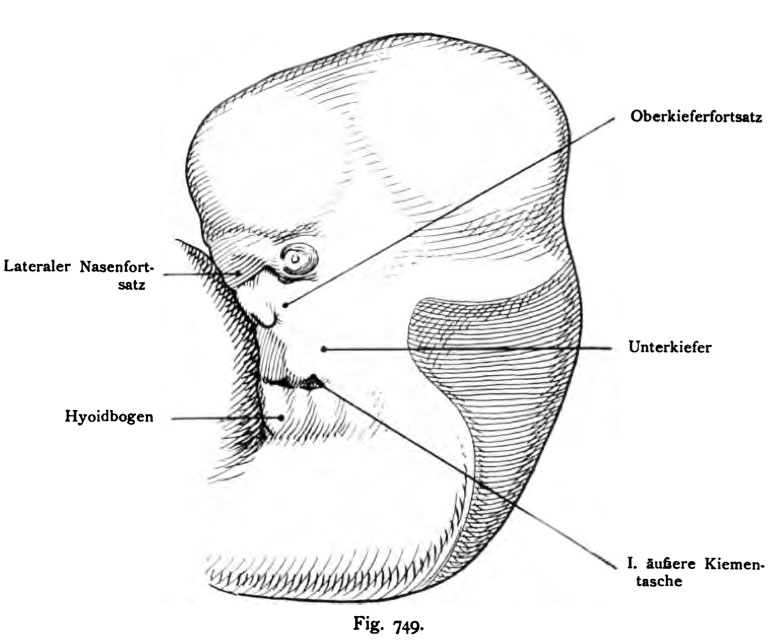
Fig. 749. Form der ersten äufteren Kiemeatasche»
in deren Umgebung sich später das äußere Ohr anlegt und deren Rinne sieh in den Meatus acusticus extemus umwandelt. Menschlicher Embryo von 11,3 mm,
Linke Seite, Profil,
(Nach Rabl.)
Die Spalte zeigt zwei Erweiterungen, eine dorsale und eine ventrale Ecke. Die naheliegenden Abschnitte des Mandibular- und des Hyoidbogens zeigen rundliche Erhebungen: die Aurikularhöcker, Colliculi auriculares.
File history
Click on a date/time to view the file as it appeared at that time.
| Date/Time | Thumbnail | Dimensions | User | Comment | |
|---|---|---|---|---|---|
| current | 12:34, 21 October 2011 | | 774 × 641 (66 KB) | S8600021 (talk | contribs) | {{Kollmann1907}} Category:Hearing Fig. 749. Form der ersten äufteren Kiemeatasche» in deren Umgebung sich später das äußere Ohr anlegt und deren Rinne sieh in den Meatus acusticus extemus umwandelt. Menschlicher Embryo von 11,3 mm, Linke |
You cannot overwrite this file.
File usage
The following page uses this file: