File:Kollmann747.jpg
Kollmann747.jpg (753 × 388 pixels, file size: 44 KB, MIME type: image/jpeg)
- This text is a Google translate computer generated translation and may contain many errors.
Images from - Atlas of the Development of Man (Volume 2)
(Handatlas der entwicklungsgeschichte des menschen)
- Kollmann Atlas 2: Gastrointestinal | Respiratory | Urogenital | Cardiovascular | Neural | Integumentary | Smell | Vision | Hearing | Kollmann Atlas 1 | Kollmann Atlas 2 | Julius Kollmann
- Links: Julius Kollman | Atlas Vol.1 | Atlas Vol.2 | Embryology History
| Historic Disclaimer - information about historic embryology pages |
|---|
 |
Reference
Kollmann JKE. Atlas of the Development of Man (Handatlas der entwicklungsgeschichte des menschen). (1907) Vol.1 and Vol. 2. Jena, Gustav Fischer. (1898).
Cite this page: Hill, M.A. (2024, April 23) Embryology Kollmann747.jpg. Retrieved from https://embryology.med.unsw.edu.au/embryology/index.php/File:Kollmann747.jpg
- © Dr Mark Hill 2024, UNSW Embryology ISBN: 978 0 7334 2609 4 - UNSW CRICOS Provider Code No. 00098G
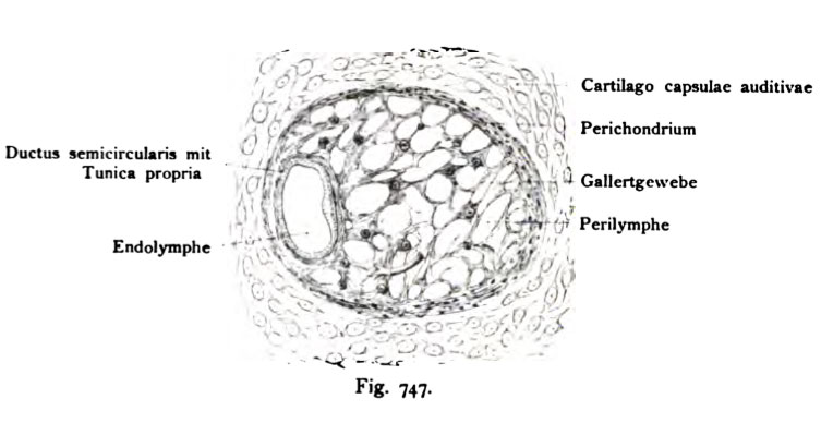
Fig. 747. Ductus semicircularis membranaceus im Ductus semicircularis
cartUagineus liesend.
Durchschnitt. (Die Umrisse sind 80 mal vergr. Die histologischen Elemente mehr.)
Menschlicher Fetus aus dem Anfang des 3. Monats.
(Anatomische Sammlung in Basel.)
Der Querschnitt des Ductus semicircularis membranaceus nimmt nur einen kleinen Teil des Querschnittes ein. Der größere Teil gehört dem Ductus semicircularis cartilagineus an, der später verknöchert. An seiner Innenwand befindet sich eine Lage von Perichondrium , das später zum Periost sich um- wandelt. Rings um den randständig verlaufenden Kanal befindet sich areoläres Bindegewebe, dessen weite Maschen mit Perilymphe gefüllt sind.
File history
Click on a date/time to view the file as it appeared at that time.
| Date/Time | Thumbnail | Dimensions | User | Comment | |
|---|---|---|---|---|---|
| current | 12:33, 21 October 2011 | | 753 × 388 (44 KB) | S8600021 (talk | contribs) | {{Kollmann1907}} Category:Hearing Fig. 747. Ductus semicircularis membranaceus im Ductus semicircularis cartUagineus liesend. Durchschnitt. (Die Umrisse sind 80 mal vergr. Die histologischen Elemente mehr.) Menschlicher Fetus aus dem Anfang de |
You cannot overwrite this file.
File usage
The following 2 pages use this file: