File:Kollmann699.jpg
Kollmann699.jpg (722 × 418 pixels, file size: 64 KB, MIME type: image/jpeg)
- This text is a Google translate computer generated translation and may contain many errors.
Images from - Atlas of the Development of Man (Volume 2)
(Handatlas der entwicklungsgeschichte des menschen)
- Kollmann Atlas 2: Gastrointestinal | Respiratory | Urogenital | Cardiovascular | Neural | Integumentary | Smell | Vision | Hearing | Kollmann Atlas 1 | Kollmann Atlas 2 | Julius Kollmann
- Links: Julius Kollman | Atlas Vol.1 | Atlas Vol.2 | Embryology History
| Historic Disclaimer - information about historic embryology pages |
|---|
 |
Reference
Kollmann JKE. Atlas of the Development of Man (Handatlas der entwicklungsgeschichte des menschen). (1907) Vol.1 and Vol. 2. Jena, Gustav Fischer. (1898).
Cite this page: Hill, M.A. (2024, April 16) Embryology Kollmann699.jpg. Retrieved from https://embryology.med.unsw.edu.au/embryology/index.php/File:Kollmann699.jpg
- © Dr Mark Hill 2024, UNSW Embryology ISBN: 978 0 7334 2609 4 - UNSW CRICOS Provider Code No. 00098G
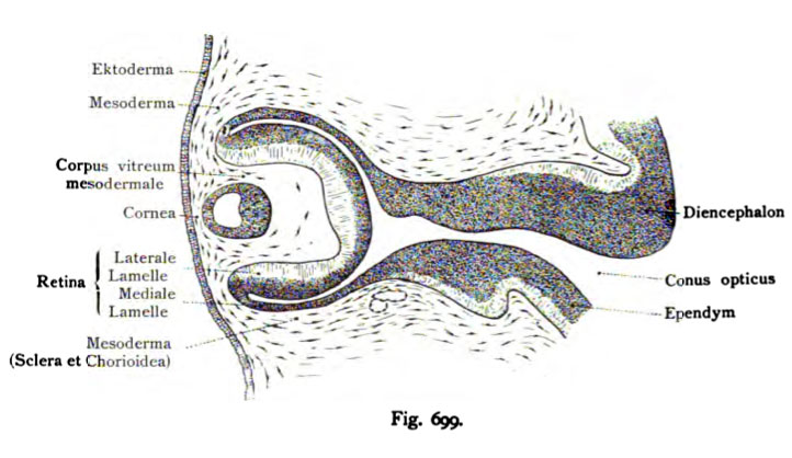
Fig. 699. Der embryonale Bulbus eines menschlichen Embryo von 10,2 mm
Länge«
(Kombiniertes Bild.) (Anatomische Sammlung in Basel.)
Die Linse hat sich jetzt von dem Ektoderm abgeschnürt, liegt aber noch sehr oberflächlich. Zwischen ihr und der lateralen Lamelle des Augenbechers existiert ein ansehnlicher Raum. Der Augen blasenstiel ist länger geworden und ist samt Augenbecher und Linse von Mesoderm umschlossen, aus dem sich die Cornea, Sclera und Chorioidea allmählich gestalten.
- Note - This image was originally uploaded as part of an undergraduate science student project and may contain inaccuracies in either description or acknowledgements. Students have been advised in writing concerning the reuse of content and may accidentally have misunderstood the original terms of use. If image reuse on this non-commercial educational site infringes your existing copyright, please contact the site editor for immediate removal.
File history
Click on a date/time to view the file as it appeared at that time.
| Date/Time | Thumbnail | Dimensions | User | Comment | |
|---|---|---|---|---|---|
| current | 10:40, 21 October 2011 | | 722 × 418 (64 KB) | S8600021 (talk | contribs) | {{Kollmann1907}} Category:Vision Fig. 699. Der embryonale Bulbus eines menschlichen Embryo von 10,2 mm Länge« (Kombiniertes Bild.) (Anatomische Sammlung in Basel.) Die Linse hat sich jetzt von dem Ektoderm abgeschnürt, liegt aber noch |
You cannot overwrite this file.
File usage
The following 3 pages use this file: