File:Kollmann697.jpg: Difference between revisions
({{Kollmann1907}} Category:Vision Fig. 697. Umwandluns der primären Augenblase des menschlichen Embryo in eine sekundäre Augenblase. (Nach His.) Die Linse hängt noch mit dem Ektoderm zusammen, die primäre Augen- blase ist gegenüber der) |
No edit summary |
||
| Line 18: | Line 18: | ||
körper seine Entwicklung gestattet. Rechts ist das Linsensäckchen nur am | körper seine Entwicklung gestattet. Rechts ist das Linsensäckchen nur am | ||
Rande getroffen, links durch die Mitte. | Rande getroffen, links durch die Mitte. | ||
{{Template:Student Image}} | |||
Revision as of 10:15, 3 October 2012
- This text is a Google translate computer generated translation and may contain many errors.
Images from - Atlas of the Development of Man (Volume 2)
(Handatlas der entwicklungsgeschichte des menschen)
- Kollmann Atlas 2: Gastrointestinal | Respiratory | Urogenital | Cardiovascular | Neural | Integumentary | Smell | Vision | Hearing | Kollmann Atlas 1 | Kollmann Atlas 2 | Julius Kollmann
- Links: Julius Kollman | Atlas Vol.1 | Atlas Vol.2 | Embryology History
| Historic Disclaimer - information about historic embryology pages |
|---|
 |
Reference
Kollmann JKE. Atlas of the Development of Man (Handatlas der entwicklungsgeschichte des menschen). (1907) Vol.1 and Vol. 2. Jena, Gustav Fischer. (1898).
Cite this page: Hill, M.A. (2024, April 19) Embryology Kollmann697.jpg. Retrieved from https://embryology.med.unsw.edu.au/embryology/index.php/File:Kollmann697.jpg
- © Dr Mark Hill 2024, UNSW Embryology ISBN: 978 0 7334 2609 4 - UNSW CRICOS Provider Code No. 00098G
Fig. 697. Umwandluns der primären Augenblase des menschlichen Embryo
in eine sekundäre Augenblase.
(Nach His.)
Die Linse hängt noch mit dem Ektoderm zusammen, die primäre Augen- blase ist gegenüber der Linsenanlage eingebuchtet. In diese Einbuchtung rückt die Linsenanlage hinein. Zwischen der Linse und der lateralen Lamelle der Augenblase ist ein schmaler Raum, der sich später vergrößert und dem Glas- körper seine Entwicklung gestattet. Rechts ist das Linsensäckchen nur am Rande getroffen, links durch die Mitte.
- Note - This image was originally uploaded as part of an undergraduate science student project and may contain inaccuracies in either description or acknowledgements. Students have been advised in writing concerning the reuse of content and may accidentally have misunderstood the original terms of use. If image reuse on this non-commercial educational site infringes your existing copyright, please contact the site editor for immediate removal.
File history
Click on a date/time to view the file as it appeared at that time.
| Date/Time | Thumbnail | Dimensions | User | Comment | |
|---|---|---|---|---|---|
| current | 10:39, 21 October 2011 | 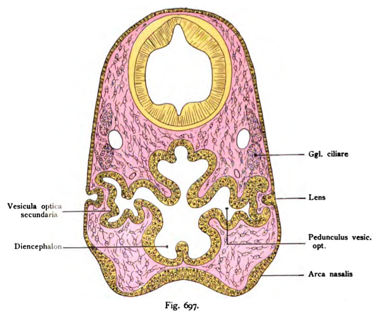 | 731 × 612 (94 KB) | S8600021 (talk | contribs) | {{Kollmann1907}} Category:Vision Fig. 697. Umwandluns der primären Augenblase des menschlichen Embryo in eine sekundäre Augenblase. (Nach His.) Die Linse hängt noch mit dem Ektoderm zusammen, die primäre Augen- blase ist gegenüber der |
You cannot overwrite this file.
File usage
The following 3 pages use this file: