File:Kollmann693.jpg: Difference between revisions
({{Kollmann1907}} Category:Vision Fig. 693. Primäre Augenblase eines menschlichen Embryo von 3,2 mm Länge. Rekonstruktion. (Nach His.) Vollbild von der rechten Seite gesehen. Das Hirnrohr ist nach Entfer- nung des Ektoderms und aller ventra) |
No edit summary |
||
| Line 15: | Line 15: | ||
bildet einen etwas abgeplatteten hohlen Vorsprung am Prosencephalon, der | bildet einen etwas abgeplatteten hohlen Vorsprung am Prosencephalon, der | ||
jetzt noch seitlich abgeht an der Berührungsgrenze von Grund- und Fltige\p\atte. | jetzt noch seitlich abgeht an der Berührungsgrenze von Grund- und Fltige\p\atte. | ||
{{Template:Student Image}} | |||
Latest revision as of 10:14, 3 October 2012
- This text is a Google translate computer generated translation and may contain many errors.
Images from - Atlas of the Development of Man (Volume 2)
(Handatlas der entwicklungsgeschichte des menschen)
- Kollmann Atlas 2: Gastrointestinal | Respiratory | Urogenital | Cardiovascular | Neural | Integumentary | Smell | Vision | Hearing | Kollmann Atlas 1 | Kollmann Atlas 2 | Julius Kollmann
- Links: Julius Kollman | Atlas Vol.1 | Atlas Vol.2 | Embryology History
| Historic Disclaimer - information about historic embryology pages |
|---|
 |
Reference
Kollmann JKE. Atlas of the Development of Man (Handatlas der entwicklungsgeschichte des menschen). (1907) Vol.1 and Vol. 2. Jena, Gustav Fischer. (1898).
Cite this page: Hill, M.A. (2024, April 19) Embryology Kollmann693.jpg. Retrieved from https://embryology.med.unsw.edu.au/embryology/index.php/File:Kollmann693.jpg
- © Dr Mark Hill 2024, UNSW Embryology ISBN: 978 0 7334 2609 4 - UNSW CRICOS Provider Code No. 00098G
Fig. 693. Primäre Augenblase eines menschlichen Embryo von 3,2 mm Länge.
Rekonstruktion. (Nach His.)
Vollbild von der rechten Seite gesehen. Das Hirnrohr ist nach Entfer- nung des Ektoderms und aller ventral liegenden Organe, wie Herz, Darmrohr usw. von links dargestellt. Die primäre Augenblase, Vesicula optica primitiva, bildet einen etwas abgeplatteten hohlen Vorsprung am Prosencephalon, der jetzt noch seitlich abgeht an der Berührungsgrenze von Grund- und Fltige\p\atte.
- Note - This image was originally uploaded as part of an undergraduate science student project and may contain inaccuracies in either description or acknowledgements. Students have been advised in writing concerning the reuse of content and may accidentally have misunderstood the original terms of use. If image reuse on this non-commercial educational site infringes your existing copyright, please contact the site editor for immediate removal.
File history
Click on a date/time to view the file as it appeared at that time.
| Date/Time | Thumbnail | Dimensions | User | Comment | |
|---|---|---|---|---|---|
| current | 10:37, 21 October 2011 | 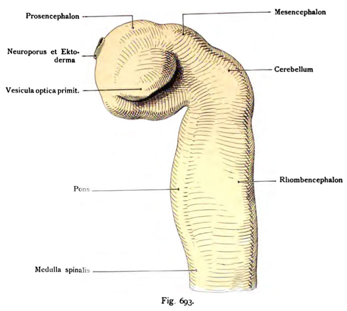 | 693 × 621 (56 KB) | S8600021 (talk | contribs) | {{Kollmann1907}} Category:Vision Fig. 693. Primäre Augenblase eines menschlichen Embryo von 3,2 mm Länge. Rekonstruktion. (Nach His.) Vollbild von der rechten Seite gesehen. Das Hirnrohr ist nach Entfer- nung des Ektoderms und aller ventra |
You cannot overwrite this file.
File usage
The following 3 pages use this file: