File:Kollmann692.jpg
Kollmann692.jpg (603 × 371 pixels, file size: 38 KB, MIME type: image/jpeg)
- This text is a Google translate computer generated translation and may contain many errors.
Images from - Atlas of the Development of Man (Volume 2)
(Handatlas der entwicklungsgeschichte des menschen)
- Kollmann Atlas 2: Gastrointestinal | Respiratory | Urogenital | Cardiovascular | Neural | Integumentary | Smell | Vision | Hearing | Kollmann Atlas 1 | Kollmann Atlas 2 | Julius Kollmann
- Links: Julius Kollman | Atlas Vol.1 | Atlas Vol.2 | Embryology History
| Historic Disclaimer - information about historic embryology pages |
|---|
 |
Reference
Kollmann JKE. Atlas of the Development of Man (Handatlas der entwicklungsgeschichte des menschen). (1907) Vol.1 and Vol. 2. Jena, Gustav Fischer. (1898).
Cite this page: Hill, M.A. (2024, April 23) Embryology Kollmann692.jpg. Retrieved from https://embryology.med.unsw.edu.au/embryology/index.php/File:Kollmann692.jpg
- © Dr Mark Hill 2024, UNSW Embryology ISBN: 978 0 7334 2609 4 - UNSW CRICOS Provider Code No. 00098G
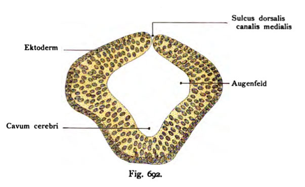
Fig. 692. Entstehung des lichtempfindenden Apparates.
Die Augengegend als eine schalenförmige Ausbuchtung des Vorderhims noch an
der Seitenwand befindlich (Augenfeld).
(Nach Heape, aus Nussbaum S. 6.)
Später wachsen diese Augenfelder zu seitlichen Divertikeln aus und stellen dann die primitive Augenblase dar, welche durch einen engen Kanal mit dem Prosencephalon und später mit dem Diencephalon zusammenhängt (Vergl. die Figg. 693 und 694.)
- Note - This image was originally uploaded as part of an undergraduate science student project and may contain inaccuracies in either description or acknowledgements. Students have been advised in writing concerning the reuse of content and may accidentally have misunderstood the original terms of use. If image reuse on this non-commercial educational site infringes your existing copyright, please contact the site editor for immediate removal.
File history
Click on a date/time to view the file as it appeared at that time.
| Date/Time | Thumbnail | Dimensions | User | Comment | |
|---|---|---|---|---|---|
| current | 10:37, 21 October 2011 | | 603 × 371 (38 KB) | S8600021 (talk | contribs) | {{Kollmann1907}} Category:Vision Fig. 692. Entstehung des lichtempfindenden Apparates. Die Augengegend als eine schalenförmige Ausbuchtung des Vorderhims noch an der Seitenwand befindlich (Augenfeld). (Nach Heape, aus Nussbaum S. 6.) Sp� |
You cannot overwrite this file.
File usage
The following 3 pages use this file: